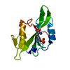
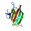
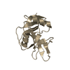
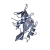
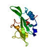
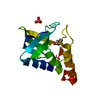
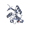
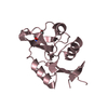
BIOMOLECULE: 1 THIS ENTRY CONTAINS THE CRYSTALLOGRAPHIC ASYMMETRIC UNIT WHICH CONSISTS OF 1 CHAIN(S) ...BIOMOLECULE: 1 THIS ENTRY CONTAINS THE CRYSTALLOGRAPHIC ASYMMETRIC UNIT WHICH CONSISTS OF 1 CHAIN(S). SEE REMARK 350 FOR INFORMATION ON GENERATING THE BIOLOGICAL MOLECULE(S). ACCORDING TO AUTHORS, INTACT HUMAN CD23 IS THOUGHT TO FORM A TRIMER, HOWEVER, THE EXACT RELATIONSHIP OF THE LECTIN DOMAINS IN THE MULTIMER IS NOT CURRENTLY KNOWN
Intact human CD23 is thought to form a trimer, although it is possible that it forms a tetramer. The exact relationship of the monomer lectin domains to each other is not currently known.
Resolution: 1.3→38.63 Å / Cor.coef. Fo:Fc: 0.963 / Cor.coef. Fo:Fc free: 0.955 / SU B: 1.37 / SU ML: 0.027 / Cross valid method: THROUGHOUT / ESU R: 0.057 / ESU R Free: 0.054 / Stereochemistry target values: MAXIMUM LIKELIHOOD / Details: HYDROGENS HAVE BEEN ADDED IN THE RIDING POSITIONS
Rfactor
Num. reflection
% reflection
Selection details
Rfree
0.20045
1561
5.1 %
THIN SHELLS
Rwork
0.16785
-
-
-
all
0.16953
28870
-
-
obs
0.16953
28870
99.37 %
-
Solvent computation
Ion probe radii: 0.8 Å / Shrinkage radii: 0.8 Å / VDW probe radii: 1.2 Å / Solvent model: MASK
Displacement parameters
Biso mean: 15.865 Å2
Baniso -1
Baniso -2
Baniso -3
1-
0.52 Å2
0 Å2
0 Å2
2-
-
-0.56 Å2
0 Å2
3-
-
-
0.04 Å2
Refinement step
Cycle: LAST / Resolution: 1.3→38.63 Å
Protein
Nucleic acid
Ligand
Solvent
Total
Num. atoms
1001
0
1
157
1159
Refine LS restraints
Refine-ID
Type
Dev ideal
Dev ideal target
Number
X-RAY DIFFRACTION
r_bond_refined_d
0.017
0.021
1058
X-RAY DIFFRACTION
r_bond_other_d
0.002
0.02
894
X-RAY DIFFRACTION
r_angle_refined_deg
1.658
1.906
1435
X-RAY DIFFRACTION
r_angle_other_deg
2.527
3
2085
X-RAY DIFFRACTION
r_dihedral_angle_1_deg
6.723
5
128
X-RAY DIFFRACTION
r_dihedral_angle_2_deg
30.961
23.091
55
X-RAY DIFFRACTION
r_dihedral_angle_3_deg
11.692
15
173
X-RAY DIFFRACTION
r_dihedral_angle_4_deg
17.542
15
9
X-RAY DIFFRACTION
r_chiral_restr
0.099
0.2
139
X-RAY DIFFRACTION
r_gen_planes_refined
0.007
0.02
1193
X-RAY DIFFRACTION
r_gen_planes_other
0.001
0.02
241
X-RAY DIFFRACTION
r_nbd_refined
0.236
0.2
213
X-RAY DIFFRACTION
r_nbd_other
0.222
0.2
951
X-RAY DIFFRACTION
r_nbtor_refined
0.194
0.2
501
X-RAY DIFFRACTION
r_nbtor_other
0.087
0.2
597
X-RAY DIFFRACTION
r_xyhbond_nbd_refined
0.203
0.2
102
X-RAY DIFFRACTION
r_xyhbond_nbd_other
0.251
0.2
1
X-RAY DIFFRACTION
r_metal_ion_refined
0.031
0.2
3
X-RAY DIFFRACTION
r_symmetry_vdw_refined
0.227
0.2
11
X-RAY DIFFRACTION
r_symmetry_vdw_other
0.4
0.2
35
X-RAY DIFFRACTION
r_symmetry_hbond_refined
0.237
0.2
13
X-RAY DIFFRACTION
r_mcbond_it
4.997
3
643
X-RAY DIFFRACTION
r_mcbond_other
3.363
3
264
X-RAY DIFFRACTION
r_mcangle_it
5.828
4
994
X-RAY DIFFRACTION
r_scbond_it
6.069
3
502
X-RAY DIFFRACTION
r_scangle_it
7.473
4
438
X-RAY DIFFRACTION
r_rigid_bond_restr
3.762
3
2315
X-RAY DIFFRACTION
r_sphericity_free
18.203
3
155
X-RAY DIFFRACTION
r_sphericity_bonded
7.9
3
1917
LS refinement shell
Resolution: 1.3→1.334 Å / Total num. of bins used: 20
Rfactor
Num. reflection
% reflection
Rfree
0.39
77
-
Rwork
0.292
2134
-
obs
-
2134
100 %
+
About Yorodumi
-
News
-
Feb 9, 2022. New format data for meta-information of EMDB entries
New format data for meta-information of EMDB entries
Version 3 of the EMDB header file is now the official format.
The previous official version 1.9 will be removed from the archive.
In the structure databanks used in Yorodumi, some data are registered as the other names, "COVID-19 virus" and "2019-nCoV". Here are the details of the virus and the list of structure data.
Jan 31, 2019. EMDB accession codes are about to change! (news from PDBe EMDB page)
EMDB accession codes are about to change! (news from PDBe EMDB page)
The allocation of 4 digits for EMDB accession codes will soon come to an end. Whilst these codes will remain in use, new EMDB accession codes will include an additional digit and will expand incrementally as the available range of codes is exhausted. The current 4-digit format prefixed with “EMD-” (i.e. EMD-XXXX) will advance to a 5-digit format (i.e. EMD-XXXXX), and so on. It is currently estimated that the 4-digit codes will be depleted around Spring 2019, at which point the 5-digit format will come into force.
The EM Navigator/Yorodumi systems omit the EMD- prefix.
Related info.:Q: What is EMD? / ID/Accession-code notation in Yorodumi/EM Navigator
Yorodumi is a browser for structure data from EMDB, PDB, SASBDB, etc.
This page is also the successor to EM Navigator detail page, and also detail information page/front-end page for Omokage search.
The word "yorodu" (or yorozu) is an old Japanese word meaning "ten thousand". "mi" (miru) is to see.
Related info.:EMDB / PDB / SASBDB / Comparison of 3 databanks / Yorodumi Search / Aug 31, 2016. New EM Navigator & Yorodumi / Yorodumi Papers / Jmol/JSmol / Function and homology information / Changes in new EM Navigator and Yorodumi
 Movie
Movie Controller


 Open data
 Basic information
 Components
 Keywords
 Function and homology information
 Homo sapiens (human)
 X-RAY DIFFRACTION /
  Authors
 Citation
 Structure visualization
 Downloads & links
 Other downloads


 PDBj
PDBj


 Assembly

 Spodoptera frugiperda (fall armyworm) / Strain (production host): Sf+ / References: UniProt: P06734
 Mass: 40.078 Da / Num. of mol.: 1 / Source method: obtained synthetically / Formula: Ca
 Mass: 18.015 Da / Num. of mol.: 157 / Source method: isolated from a natural source / Formula: H2O
 Sample preparation
 / Beamline: 32-ID / Wavelength: 1.02336 Å
 Processing