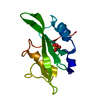
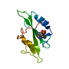
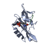
[English] 日本語
Yorodumi
- PDB-4f5b: Triple mutant Src SH2 domain bound to phosphotyrosine -

+
Open data


ID or keywords:

Loading...

-
Basic information

Entry
Database: PDB / ID: 4f5b
TitleTriple mutant Src SH2 domain bound to phosphotyrosine
ComponentsProto-oncogene tyrosine-protein kinase Src
KeywordsPROTEIN BINDING / SH2 domain / Cell signalling / phosphotyrosine
Function / homology
Function and homology information


regulation of caveolin-mediated endocytosis / regulation of toll-like receptor 3 signaling pathway / regulation of cell projection assembly / positive regulation of platelet-derived growth factor receptor-beta signaling pathway / cellular response to prolactin / positive regulation of male germ cell proliferation / dendritic filopodium / negative regulation of telomere maintenance / response to mineralocorticoid / Regulation of commissural axon pathfinding by SLIT and ROBO ...regulation of caveolin-mediated endocytosis / regulation of toll-like receptor 3 signaling pathway / regulation of cell projection assembly / positive regulation of platelet-derived growth factor receptor-beta signaling pathway / cellular response to prolactin / positive regulation of male germ cell proliferation / dendritic filopodium / negative regulation of telomere maintenance / response to mineralocorticoid / Regulation of commissural axon pathfinding by SLIT and ROBO / positive regulation of dephosphorylation / regulation of epithelial cell migration / ERBB2 signaling pathway / positive regulation of protein transport / Regulation of gap junction activity / cellular response to progesterone stimulus / BMP receptor binding / negative regulation of focal adhesion assembly / positive regulation of protein processing / skeletal muscle cell proliferation / positive regulation of integrin activation / Activated NTRK2 signals through FYN / Netrin mediated repulsion signals / regulation of intracellular estrogen receptor signaling pathway / intestinal epithelial cell development / regulation of vascular permeability / negative regulation of neutrophil activation / positive regulation of glucose metabolic process / transcytosis / osteoclast development / Activated NTRK3 signals through PI3K / connexin binding / focal adhesion assembly / cellular response to fluid shear stress / adherens junction organization / signal complex assembly / positive regulation of small GTPase mediated signal transduction / Co-stimulation by CD28 / branching involved in mammary gland duct morphogenesis / response to acidic pH / positive regulation of Ras protein signal transduction / Regulation of RUNX1 Expression and Activity / DCC mediated attractive signaling / positive regulation of podosome assembly / EPH-Ephrin signaling / regulation of bone resorption / positive regulation of lamellipodium morphogenesis / Ephrin signaling / myoblast proliferation / Signal regulatory protein family interactions / cellular response to peptide hormone stimulus / negative regulation of mitochondrial depolarization / podosome / odontogenesis / MET activates PTK2 signaling / cellular response to fatty acid / Regulation of KIT signaling / postsynaptic specialization, intracellular component / regulation of early endosome to late endosome transport / Signaling by ALK / leukocyte migration / phospholipase activator activity / Co-inhibition by CTLA4 / oogenesis / GP1b-IX-V activation signalling / Receptor Mediated Mitophagy / EPHA-mediated growth cone collapse / interleukin-6-mediated signaling pathway / p130Cas linkage to MAPK signaling for integrins / positive regulation of smooth muscle cell migration / DNA biosynthetic process / positive regulation of Notch signaling pathway / Signaling by EGFR / stress fiber assembly / RUNX2 regulates osteoblast differentiation / stimulatory C-type lectin receptor signaling pathway / negative regulation of intrinsic apoptotic signaling pathway / uterus development / regulation of cell-cell adhesion / Fc-gamma receptor signaling pathway involved in phagocytosis / phospholipase binding / PECAM1 interactions / Recycling pathway of L1 / regulation of heart rate by cardiac conduction / neurotrophin TRK receptor signaling pathway / GRB2:SOS provides linkage to MAPK signaling for Integrins / dendritic growth cone / protein tyrosine kinase activator activity / RHOU GTPase cycle / platelet-derived growth factor receptor signaling pathway / negative regulation of anoikis / RET signaling / Long-term potentiation / FCGR activation / positive regulation of epithelial cell migration / EPH-ephrin mediated repulsion of cells / ephrin receptor signaling pathway / GAB1 signalosome / positive regulation of protein serine/threonine kinase activity / negative regulation of hippo signaling
Similarity search - Function
SH2 domain / SHC Adaptor Protein / : / SH3 domain / SH2 domain / Src homology 2 (SH2) domain profile. / Src homology 2 domains / SH2 domain / SH2 domain superfamily / Src homology 3 domains ...SH2 domain / SHC Adaptor Protein / : / SH3 domain / SH2 domain / Src homology 2 (SH2) domain profile. / Src homology 2 domains / SH2 domain / SH2 domain superfamily / Src homology 3 domains / SH3-like domain superfamily / Src homology 3 (SH3) domain profile. / SH3 domain / Tyrosine-protein kinase, catalytic domain / Tyrosine kinase, catalytic domain / Tyrosine protein kinases specific active-site signature. / Tyrosine-protein kinase, active site / Serine-threonine/tyrosine-protein kinase, catalytic domain / Protein tyrosine and serine/threonine kinase / Protein kinase, ATP binding site / Protein kinases ATP-binding region signature. / Protein kinase domain profile. / Protein kinase domain / Protein kinase-like domain superfamily / 2-Layer Sandwich / Alpha Beta
Similarity search - Domain/homology
O-PHOSPHOTYROSINE / Proto-oncogene tyrosine-protein kinase Src
Similarity search - Component
Biological speciesHomo sapiens (human)
MethodX-RAY DIFFRACTION / MOLECULAR REPLACEMENT / molecular replacement / Resolution: 1.57 Å
AuthorsKaneko, T. / Huang, H. / Cao, X. / Li, C. / Voss, C. / Sidhu, S.S. / Li, S.S.
CitationJournal: Sci.Signal. / Year: 2012
Title: Superbinder SH2 Domains Act as Antagonists of Cell Signaling.
Authors: Kaneko, T. / Huang, H. / Cao, X. / Li, X. / Li, C. / Voss, C. / Sidhu, S.S. / Li, S.S.
History
DepositionMay 12, 2012Deposition site: RCSB / Processing site: RCSB
Revision 1.0Oct 3, 2012Provider: repository / Type: Initial release
Revision 1.1Feb 28, 2024Group: Data collection / Database references / Derived calculations
Category: chem_comp_atom / chem_comp_bond ...chem_comp_atom / chem_comp_bond / database_2 / struct_ref_seq_dif / struct_site
Item: _database_2.pdbx_DOI / _database_2.pdbx_database_accession ..._database_2.pdbx_DOI / _database_2.pdbx_database_accession / _struct_ref_seq_dif.details / _struct_site.pdbx_auth_asym_id / _struct_site.pdbx_auth_comp_id / _struct_site.pdbx_auth_seq_id
Revision 1.2Mar 13, 2024Group: Source and taxonomy / Structure summary / Category: entity / pdbx_entity_src_syn / Item: _entity.details

-
Structure visualization

Structure viewerMolecule:
MolmilJmol/JSmol

Downloads & links

-
Assembly

Deposited unit
A: Proto-oncogene tyrosine-protein kinase Src
hetero molecules


Theoretical massNumber of molelcules
Total (without water)12,9602
Polymers12,6991
Non-polymers2611
Water2,216123
1


  • Idetical with deposited unit
  • defined by author&software
TypeNameSymmetry operationNumber
identity operation1_555x,y,z1
MethodPISA
Unit cell
Length a, b, c (Å)67.600, 67.600, 46.800
Angle α, β, γ (deg.)90.000, 90.000, 120.000
Int Tables number169
Space group name H-MP61
Detailsbiological unit is the same as asym.

-
Components

#1: Protein Proto-oncogene tyrosine-protein kinase Src / Proto-oncogene c-Src / pp60c-src / p60-Src


Mass: 12699.316 Da / Num. of mol.: 1 / Fragment: SH2 domain / Mutation: T183V/C188A/K206L
Source method: isolated from a genetically manipulated source
Source: (gene. exp.) Homo sapiens (human) / Gene: SRC, SRC1 / Production host: Escherichia coli (E. coli) / Strain (production host): BL21(DE3) / References: UniProt: P12931
#2: Chemical ChemComp-PTR / O-PHOSPHOTYROSINE / PHOSPHONOTYROSINE


Type: L-peptide linking / Mass: 261.168 Da / Num. of mol.: 1 / Source method: obtained synthetically / Formula: C9H12NO6P / Details: phosphorylated tyrosine
#3: Water ChemComp-HOH / water


Mass: 18.015 Da / Num. of mol.: 123 / Source method: isolated from a natural source / Formula: H2O

-
Experimental details

-
Experiment

ExperimentMethod: X-RAY DIFFRACTION / Number of used crystals: 1

-
Sample preparation

CrystalDensity Matthews: 2.43 Å3/Da / Density % sol: 49.4 %
Crystal growTemperature: 277 K / Method: vapor diffusion, sitting drop / pH: 7.6
Details: 0.1 M Tris-HCl, 18% PEG6000, 0.2 M lithium chloride, pH 7.6, vapor diffusion, sitting drop, temperature 277K

-
Data collection

DiffractionMean temperature: 114 K
Diffraction sourceSource: ROTATING ANODE / Type: RIGAKU RUH3R / Wavelength: 1.5418 Å
DetectorType: MAR scanner 345 mm plate / Detector: IMAGE PLATE / Date: Oct 26, 2011
RadiationMonochromator: Graded multilayer (osmic) / Protocol: SINGLE WAVELENGTH / Monochromatic (M) / Laue (L): M / Scattering type: x-ray
Radiation wavelengthWavelength: 1.5418 Å / Relative weight: 1
ReflectionResolution: 1.57→19.514 Å / Num. all: 15470 / Num. obs: 15470 / % possible obs: 90.2 % / Observed criterion σ(F): 0 / Observed criterion σ(I): 0 / Redundancy: 8.2 % / Rsym value: 0.045 / Net I/σ(I): 30.2
Reflection shell

Diffraction-ID: 1

Resolution (Å)Redundancy (%)Rmerge(I) obsMean I/σ(I) obsNum. measured allNum. unique allRsym value% possible all
1.57-1.6580.2662.91993224980.266100
1.65-1.768.10.1714.41724621250.17190.4
1.76-1.888.20.1086.81836022370.108100
1.88-2.0380.0867.11142414290.08670.3
2.03-2.228.40.05131436517070.0589.3
2.22-2.488.30.04413.81243314900.04486.5
2.48-2.878.60.03517.51181013720.03590
2.87-3.518.70.035151061712270.03595
3.51-4.968.20.0414.970248540.0484.3
4.96-20.0047.60.04513.140505310.04592.3

-
Phasing

PhasingMethod: molecular replacement
Phasing MR
Highest resolutionLowest resolution
Rotation1.87 Å22.09 Å
Translation1.87 Å22.09 Å

-
Processing

Software
NameVersionClassificationNB
MOSFLMdata reduction
SCALA3.3.16data scaling
MOLREPphasing
REFMACrefinement
PDB_EXTRACT3.11data extraction
MAR345dtbdata collection
RefinementMethod to determine structure: MOLECULAR REPLACEMENT / Resolution: 1.57→19.51 Å / Cor.coef. Fo:Fc: 0.957 / Cor.coef. Fo:Fc free: 0.947 / WRfactor Rfree: 0.2377 / WRfactor Rwork: 0.2035 / Occupancy max: 1 / Occupancy min: 1 / FOM work R set: 0.8507 / SU B: 3.504 / SU ML: 0.057 / SU R Cruickshank DPI: 0.1005 / SU Rfree: 0.0995 / Cross valid method: THROUGHOUT / σ(F): 0 / ESU R: 0.1 / ESU R Free: 0.099 / Stereochemistry target values: MAXIMUM LIKELIHOOD
Details: HYDROGENS HAVE BEEN ADDED IN THE RIDING POSITIONS U VALUES : WITH TLS ADDED
RfactorNum. reflection% reflectionSelection details
Rfree0.2303 724 4.7 %RANDOM
Rwork0.1981 ---
all0.1996 15437 --
obs0.1996 15437 90.15 %-
Solvent computationIon probe radii: 0.8 Å / Shrinkage radii: 0.8 Å / VDW probe radii: 1.4 Å / Solvent model: MASK
Displacement parametersBiso max: 51.56 Å2 / Biso mean: 24.6385 Å2 / Biso min: 11 Å2
Baniso -1Baniso -2Baniso -3
1-0.47 Å20.23 Å20 Å2
2--0.47 Å20 Å2
3----0.7 Å2
Refinement stepCycle: LAST / Resolution: 1.57→19.51 Å
ProteinNucleic acidLigandSolventTotal
Num. atoms867 0 17 123 1007
Refine LS restraints
Refine-IDTypeDev idealDev ideal targetNumber
X-RAY DIFFRACTIONr_bond_refined_d0.0120.021901
X-RAY DIFFRACTIONr_angle_refined_deg1.4411.9581217
X-RAY DIFFRACTIONr_dihedral_angle_1_deg6.5365107
X-RAY DIFFRACTIONr_dihedral_angle_2_deg26.28922.88945
X-RAY DIFFRACTIONr_dihedral_angle_3_deg11.89815154
X-RAY DIFFRACTIONr_dihedral_angle_4_deg10.776158
X-RAY DIFFRACTIONr_chiral_restr0.0940.2132
X-RAY DIFFRACTIONr_gen_planes_refined0.0070.02683
X-RAY DIFFRACTIONr_mcbond_it0.8331.5539
X-RAY DIFFRACTIONr_mcangle_it1.5012864
X-RAY DIFFRACTIONr_scbond_it2.43362
X-RAY DIFFRACTIONr_scangle_it3.8324.5353
LS refinement shellResolution: 1.57→1.611 Å / Total num. of bins used: 20
RfactorNum. reflection% reflection
Rfree0.3 59 -
Rwork0.298 1219 -
all-1278 -
obs--100 %
Refinement TLS params.Method: refined / Origin x: -8.946 Å / Origin y: 23.559 Å / Origin z: -0.158 Å
111213212223313233
T0.096 Å20.0059 Å20.0117 Å2-0.1006 Å20.0086 Å2--0.1439 Å2
L2.0918 °2-1.2259 °20.0742 °2-1.428 °20.2568 °2--1.3051 °2
S0.0347 Å °0.0102 Å °-0.0437 Å °-0.0841 Å °-0.023 Å °0.003 Å °-0.1222 Å °-0.0054 Å °-0.0117 Å °
Refinement TLS group
IDRefine-IDRefine TLS-IDAuth asym-IDAuth seq-ID
1X-RAY DIFFRACTION1A1 - 1000
2X-RAY DIFFRACTION1A1 - 1000

+
About Yorodumi

-
News

-
Feb 9, 2022. New format data for meta-information of EMDB entries

New format data for meta-information of EMDB entries

  • Version 3 of the EMDB header file is now the official format.
  • The previous official version 1.9 will be removed from the archive.

Related info.:EMDB header

External links:wwPDB to switch to version 3 of the EMDB data model

-
Aug 12, 2020. Covid-19 info

Covid-19 info

URL: https://pdbj.org/emnavi/covid19.php

New page: Covid-19 featured information page in EM Navigator.

Related info.:Covid-19 info / Mar 5, 2020. Novel coronavirus structure data

+
Mar 5, 2020. Novel coronavirus structure data

Novel coronavirus structure data

Related info.:Yorodumi Speices / Aug 12, 2020. Covid-19 info

External links:COVID-19 featured content - PDBj / Molecule of the Month (242):Coronavirus Proteases

+
Jan 31, 2019. EMDB accession codes are about to change! (news from PDBe EMDB page)

EMDB accession codes are about to change! (news from PDBe EMDB page)

  • The allocation of 4 digits for EMDB accession codes will soon come to an end. Whilst these codes will remain in use, new EMDB accession codes will include an additional digit and will expand incrementally as the available range of codes is exhausted. The current 4-digit format prefixed with “EMD-” (i.e. EMD-XXXX) will advance to a 5-digit format (i.e. EMD-XXXXX), and so on. It is currently estimated that the 4-digit codes will be depleted around Spring 2019, at which point the 5-digit format will come into force.
  • The EM Navigator/Yorodumi systems omit the EMD- prefix.

Related info.:Q: What is EMD? / ID/Accession-code notation in Yorodumi/EM Navigator

External links:EMDB Accession Codes are Changing Soon! / Contact to PDBj

+
Jul 12, 2017. Major update of PDB

Major update of PDB

  • wwPDB released updated PDB data conforming to the new PDBx/mmCIF dictionary.
  • This is a major update changing the version number from 4 to 5, and with Remediation, in which all the entries are updated.
  • In this update, many items about electron microscopy experimental information are reorganized (e.g. em_software).
  • Now, EM Navigator and Yorodumi are based on the updated data.

External links:wwPDB Remediation / Enriched Model Files Conforming to OneDep Data Standards Now Available in the PDB FTP Archive

-
Yorodumi

Thousand views of thousand structures

  • Yorodumi is a browser for structure data from EMDB, PDB, SASBDB, etc.
  • This page is also the successor to EM Navigator detail page, and also detail information page/front-end page for Omokage search.
  • The word "yorodu" (or yorozu) is an old Japanese word meaning "ten thousand". "mi" (miru) is to see.

Related info.:EMDB / PDB / SASBDB / Comparison of 3 databanks / Yorodumi Search / Aug 31, 2016. New EM Navigator & Yorodumi / Yorodumi Papers / Jmol/JSmol / Function and homology information / Changes in new EM Navigator and Yorodumi

Read more