 Movie
Movie Controller

[English] 日本語
 Yorodumi
- PDB-6ps8: XFEL MT1R structure by ligand exchange from agomelatine to 2-phen... -
+ Open data

- Basic information
| Entry | Database: PDB / ID: 6ps8 | ||||||
|---|---|---|---|---|---|---|---|
| Title | XFEL MT1R structure by ligand exchange from agomelatine to 2-phenylmelatonin. | ||||||
 Components Components | Fusion protein of Melatonin receptor type 1A and GlgA glycogen synthase | ||||||
 Keywords Keywords | MEMBRANE PROTEIN / GPCR / COMPLEX-LCP method / SBDD / drug design / XFEL / LCP-SFX / Ligand Exchange / Agomelatine / 2-phenyl melatonin / MT1 | ||||||
| Function / homology |  Function and homology information Function and homology informationmelatonin receptor activity / alpha-1,4-glucan glucosyltransferase (UDP-glucose donor) activity / Class A/1 (Rhodopsin-like receptors) / mating behavior / hormone binding / G protein-coupled receptor signaling pathway, coupled to cyclic nucleotide second messenger / circadian rhythm / G protein-coupled receptor activity / adenylate cyclase-inhibiting G protein-coupled receptor signaling pathway / G alpha (i) signalling events ...melatonin receptor activity / alpha-1,4-glucan glucosyltransferase (UDP-glucose donor) activity / Class A/1 (Rhodopsin-like receptors) / mating behavior / hormone binding / G protein-coupled receptor signaling pathway, coupled to cyclic nucleotide second messenger / circadian rhythm / G protein-coupled receptor activity / adenylate cyclase-inhibiting G protein-coupled receptor signaling pathway / G alpha (i) signalling events / signaling receptor complex / G protein-coupled receptor signaling pathway / plasma membrane Similarity search - Function | ||||||
| Biological species |  Homo sapiens (human) Homo sapiens (human)  Pyrococcus abyssi (archaea) Pyrococcus abyssi (archaea) | ||||||
| Method |  X-RAY DIFFRACTION / FREE ELECTRON LASER / MOLECULAR REPLACEMENT / molecular replacement / Resolution: 3.3 Å X-RAY DIFFRACTION / FREE ELECTRON LASER / MOLECULAR REPLACEMENT / molecular replacement / Resolution: 3.3 Å | ||||||
 Authors Authors | Ishchenko, A. / Stauch, B. / Han, G.W. / Batyuk, A. / Shiriaeva, A. / Li, C. / Zatsepin, N.A. / Weierstall, U. / Liu, W. / Nango, E. ...Ishchenko, A. / Stauch, B. / Han, G.W. / Batyuk, A. / Shiriaeva, A. / Li, C. / Zatsepin, N.A. / Weierstall, U. / Liu, W. / Nango, E. / Nakane, T. / Tanaka, R. / Tono, K. / Joti, Y. / Iwata, S. / Moraes, I. / Gati, C. / Cherezov, C. | ||||||
| Funding support |  United States, 1items United States, 1items
| ||||||
 Citation Citation | Journal: Iucrj / Year: 2019 Title: Toward G protein-coupled receptor structure-based drug design using X-ray lasers. Authors: Ishchenko, A. / Stauch, B. / Han, G.W. / Batyuk, A. / Shiriaeva, A. / Li, C. / Zatsepin, N. / Weierstall, U. / Liu, W. / Nango, E. / Nakane, T. / Tanaka, R. / Tono, K. / Joti, Y. / Iwata, S. ...Authors: Ishchenko, A. / Stauch, B. / Han, G.W. / Batyuk, A. / Shiriaeva, A. / Li, C. / Zatsepin, N. / Weierstall, U. / Liu, W. / Nango, E. / Nakane, T. / Tanaka, R. / Tono, K. / Joti, Y. / Iwata, S. / Moraes, I. / Gati, C. / Cherezov, V. | ||||||
| History |
|
- Structure visualization
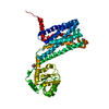
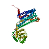
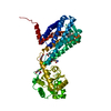
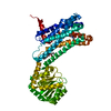
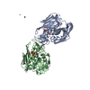
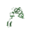
| Structure viewer | Molecule: MolmilJmol/JSmol |
|---|
- Downloads & links
-Download
| PDBx/mmCIF format | 6ps8.cif.gz | 203.4 KB | Display | PDBx/mmCIF format |
|---|---|---|---|---|
| PDB format | pdb6ps8.ent.gz | 161.1 KB | Display | PDB format |
| PDBx/mmJSON format | 6ps8.json.gz | Tree view | PDBx/mmJSON format | |
| Others |  Other downloads Other downloads |
-Validation report
| Arichive directory | https://data.pdbj.org/pub/pdb/validation_reports/ps/6ps8ftp://data.pdbj.org/pub/pdb/validation_reports/ps/6ps8 | HTTPS FTP |
|---|
-Related structure data
| Related structure data |  6przC  6ps0C  6ps1C  6ps2C  6ps3C  6ps4C  6ps5C  6ps6C  6ps7C  6me5S S: Starting model for refinement C: citing same article ( |
|---|---|
| Similar structure data |

-Links
 PDBj
PDBj


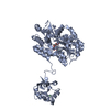
- Assembly
| Deposited unit | 
| ||||||||
|---|---|---|---|---|---|---|---|---|---|
| 1 |
| ||||||||
| Unit cell |
| ||||||||
| Details | AUTHORS STATE THAT THE BIOLOGICAL UNIT IS UNKNOWN |
-Components
| #1: Protein | Mass: 56597.355 Da / Num. of mol.: 1 Source method: isolated from a genetically manipulated source Source: (gene. exp.) Homo sapiens (human), (gene. exp.) Pyrococcus abyssi (strain GE5 / Orsay) (archaea)Gene: MTNR1A, PAB2292 / Strain: GE5 / Orsay / Production host:   Spodoptera frugiperda (fall armyworm) / References: UniProt: P48039, UniProt: Q9V2J8 Spodoptera frugiperda (fall armyworm) / References: UniProt: P48039, UniProt: Q9V2J8 |
|---|---|
| #2: Chemical | ChemComp-JEY /   Mass: 308.374 Da / Num. of mol.: 1 / Source method: obtained synthetically / Formula: C19H20N2O2 / Feature type: SUBJECT OF INVESTIGATION Mass: 308.374 Da / Num. of mol.: 1 / Source method: obtained synthetically / Formula: C19H20N2O2 / Feature type: SUBJECT OF INVESTIGATION |
| Has ligand of interest | Y |
-Experimental details
-Experiment
| Experiment | Method: X-RAY DIFFRACTION / Number of used crystals: 1 |
|---|
- Sample preparation
| Crystal | Density Matthews: 4.04 Å3/Da / Density % sol: 69.58 % |
|---|---|
| Crystal grow | Temperature: 293 K / Method: lipidic cubic phase Details: 60-100 mM potassium phosphate monobasic, 100 mM HEPES pH 7.0, 32-35% PEG 400, 1 mM of target ligand 2-phenylmelatonin, 2.5% DMSO, 1.5% propan-2-ol. |
-Data collection
| Diffraction | Mean temperature: 293 K / Serial crystal experiment: Y |
|---|---|
| Diffraction source | Source: FREE ELECTRON LASER / Site: SLAC LCLS / Beamline: CXI / Wavelength: 1.33 Å |
| Detector | Type: CS-PAD CXI-1 / Detector: PIXEL / Date: Nov 5, 2018 |
| Radiation | Protocol: SINGLE WAVELENGTH / Monochromatic (M) / Laue (L): M / Scattering type: x-ray |
| Radiation wavelength | Wavelength: 1.33 Å / Relative weight: 1 |
| Reflection | Resolution: 3.3→30 Å / Num. obs: 14561 / % possible obs: 100 % / Redundancy: 1663 % / CC1/2: 0.998 / R split: 0.119 / Net I/σ(I): 8.5 |
| Reflection shell | Resolution: 3.3→3.39 Å / Num. unique obs: 1109 / CC1/2: 0.163 / R split: 3.44 |
| Serial crystallography measurement | Collection time total: 2.27 hours / Focal spot size: 1.5 µm2 / Pulse duration: 35 fsec. / Pulse photon energy: 9.5 keV / XFEL pulse repetition rate: 120 Hz |
| Serial crystallography sample delivery | Method: injection |
| Serial crystallography sample delivery injection | Flow rate: 0.2 µL/min / Injector diameter: 50 µm |
| Serial crystallography data reduction | Crystal hits: 87453 / Frames indexed: 65260 / Frames total: 977748 |
-Phasing
| Phasing | Method: molecular replacement |
|---|
- Processing
| Software |
| |||||||||||||||||||||||||||||||||||||||||||||||||||||||||||||||||||||||||||
|---|---|---|---|---|---|---|---|---|---|---|---|---|---|---|---|---|---|---|---|---|---|---|---|---|---|---|---|---|---|---|---|---|---|---|---|---|---|---|---|---|---|---|---|---|---|---|---|---|---|---|---|---|---|---|---|---|---|---|---|---|---|---|---|---|---|---|---|---|---|---|---|---|---|---|---|---|
| Refinement | Method to determine structure: MOLECULAR REPLACEMENT Starting model: 6me5 Resolution: 3.3→30 Å / Cor.coef. Fo:Fc: 0.916 / Cor.coef. Fo:Fc free: 0.9 / SU B: 83.123 / SU ML: 0.542 / Cross valid method: THROUGHOUT / σ(F): 0 / ESU R Free: 0.533 Details: HYDROGENS HAVE BEEN ADDED IN THE RIDING POSITIONS U VALUES : WITH TLS ADDED
| |||||||||||||||||||||||||||||||||||||||||||||||||||||||||||||||||||||||||||
| Solvent computation | Ion probe radii: 0.8 Å / Shrinkage radii: 0.8 Å / VDW probe radii: 1.2 Å | |||||||||||||||||||||||||||||||||||||||||||||||||||||||||||||||||||||||||||
| Displacement parameters | Biso max: 201.26 Å2 / Biso mean: 114.238 Å2 / Biso min: 89.21 Å2
| |||||||||||||||||||||||||||||||||||||||||||||||||||||||||||||||||||||||||||
| Refinement step | Cycle: final / Resolution: 3.3→30 Å
| |||||||||||||||||||||||||||||||||||||||||||||||||||||||||||||||||||||||||||
| Refine LS restraints |
| |||||||||||||||||||||||||||||||||||||||||||||||||||||||||||||||||||||||||||
| LS refinement shell | Resolution: 3.3→3.385 Å / Rfactor Rfree error: 0 / Total num. of bins used: 20
| |||||||||||||||||||||||||||||||||||||||||||||||||||||||||||||||||||||||||||
| Refinement TLS params. | Method: refined / Refine-ID: X-RAY DIFFRACTION
| |||||||||||||||||||||||||||||||||||||||||||||||||||||||||||||||||||||||||||
| Refinement TLS group |
|
