 ムービー
ムービー コントローラー


+ データを開く

- 基本情報
基本情報
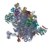
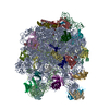
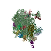
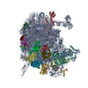
| 登録情報 | データベース: PDB / ID: 6pj6 | ||||||||||||
|---|---|---|---|---|---|---|---|---|---|---|---|---|---|
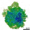
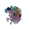
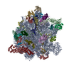
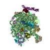
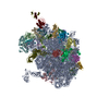
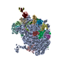
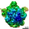
| タイトル | High resolution cryo-EM structure of E.coli 50S | ||||||||||||
 要素 要素 |
| ||||||||||||
 キーワード キーワード | RIBOSOME | ||||||||||||
| 機能・相同性 |  機能・相同性情報 機能・相同性情報large ribosomal subunit / transferase activity / 5S rRNA binding / ribosomal large subunit assembly / cytosolic large ribosomal subunit / cytoplasmic translation / tRNA binding / rRNA binding / structural constituent of ribosome / ribosome ...large ribosomal subunit / transferase activity / 5S rRNA binding / ribosomal large subunit assembly / cytosolic large ribosomal subunit / cytoplasmic translation / tRNA binding / rRNA binding / structural constituent of ribosome / ribosome / translation / ribonucleoprotein complex / cytosol / cytoplasm 類似検索 - 分子機能 | ||||||||||||
| 生物種 |  | ||||||||||||
| 手法 | 電子顕微鏡法 / 単粒子再構成法 / クライオ電子顕微鏡法 / 解像度: 2.2 Å | ||||||||||||
 データ登録者 データ登録者 | Stojkovic, V. / Myasnikov, A. / Frost, A. / Fujimori, D.G. | ||||||||||||
| 資金援助 |  米国, 3件 米国, 3件
| ||||||||||||
 引用 引用 | ジャーナル: Nucleic Acids Res / 年: 2020 タイトル: Assessment of the nucleotide modifications in the high-resolution cryo-electron microscopy structure of the Escherichia coli 50S subunit. 著者: Vanja Stojković / Alexander G Myasnikov / Iris D Young / Adam Frost / James S Fraser / Danica Galonić Fujimori / 要旨: Post-transcriptional ribosomal RNA (rRNA) modifications are present in all organisms, but their exact functional roles and positions are yet to be fully characterized. Modified nucleotides have been ...Post-transcriptional ribosomal RNA (rRNA) modifications are present in all organisms, but their exact functional roles and positions are yet to be fully characterized. Modified nucleotides have been implicated in the stabilization of RNA structure and regulation of ribosome biogenesis and protein synthesis. In some instances, rRNA modifications can confer antibiotic resistance. High-resolution ribosome structures are thus necessary for precise determination of modified nucleotides' positions, a task that has previously been accomplished by X-ray crystallography. Here, we present a cryo-electron microscopy (cryo-EM) structure of the Escherichia coli 50S subunit at an average resolution of 2.2 Å as an additional approach for mapping modification sites. Our structure confirms known modifications present in 23S rRNA and additionally allows for localization of Mg2+ ions and their coordinated water molecules. Using our cryo-EM structure as a testbed, we developed a program for assessment of cryo-EM map quality. This program can be easily used on any RNA-containing cryo-EM structure, and an associated Coot plugin allows for visualization of validated modifications, making it highly accessible. | ||||||||||||
| 履歴 |
|
- 構造の表示
構造の表示
| ムービー |
ムービービューア |
|---|---|
| 構造ビューア | 分子: MolmilJmol/JSmol |
- ダウンロードとリンク
-ダウンロード
| PDBx/mmCIF形式 | 6pj6.cif.gz | 3.2 MB | 表示 | PDBx/mmCIF形式 |
|---|---|---|---|---|
| PDB形式 | pdb6pj6.ent.gz | 表示 | PDB形式 | |
| PDBx/mmJSON形式 | 6pj6.json.gz | ツリー表示 | PDBx/mmJSON形式 | |
| その他 |  その他のダウンロード その他のダウンロード |
-検証レポート
| 文書・要旨 | 6pj6_validation.pdf.gz | 1.3 MB | 表示 | wwPDB検証レポート |
|---|---|---|---|---|
| 文書・詳細版 | 6pj6_full_validation.pdf.gz | 1.3 MB | 表示 | |
| XML形式データ | 6pj6_validation.xml.gz | 132.3 KB | 表示 | |
| CIF形式データ | 6pj6_validation.cif.gz | 250.3 KB | 表示 | |
| アーカイブディレクトリ | https://data.pdbj.org/pub/pdb/validation_reports/pj/6pj6ftp://data.pdbj.org/pub/pdb/validation_reports/pj/6pj6 | HTTPS FTP |
-関連構造データ
-リンク
 PDBj
PDBj


- 集合体
集合体
| 登録構造単位 | 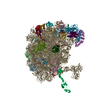
|
|---|---|
| 1 |
|
-要素
-RNA鎖 , 2種, 2分子 IJ
| #1: RNA鎖 | 分子量: 941811.562 Da / 分子数: 1 / 由来タイプ: 天然 / 由来: (天然) |
|---|---|
| #2: RNA鎖 | 分子量: 38177.762 Da / 分子数: 1 / 由来タイプ: 天然 / 由来: (天然) |
+50S ribosomal protein ... , 29種, 29分子 KLMNOPQRSTUVWXYZabcdefghijklm
-非ポリマー , 4種, 4084分子 


| #32: 化合物 | ChemComp-MG /  分子量: 24.305 Da / 分子数: 185 / 由来タイプ: 合成 / 式: Mg 分子量: 24.305 Da / 分子数: 185 / 由来タイプ: 合成 / 式: Mg#33: 化合物 | ChemComp-NA / |  分子量: 22.990 Da / 分子数: 1 / 由来タイプ: 合成 / 式: Na 分子量: 22.990 Da / 分子数: 1 / 由来タイプ: 合成 / 式: Na#34: 化合物 | ChemComp-ZN / |  分子量: 65.409 Da / 分子数: 1 / 由来タイプ: 合成 / 式: Zn 分子量: 65.409 Da / 分子数: 1 / 由来タイプ: 合成 / 式: Zn#35: 水 | ChemComp-HOH / | 分子量: 18.015 Da / 分子数: 3897 / 由来タイプ: 天然 / 式: H2O |
|---|
-実験情報
-実験
| 実験 | 手法: 電子顕微鏡法 |
|---|---|
| EM実験 | 試料の集合状態: PARTICLE / 3次元再構成法: 単粒子再構成法 |
- 試料調製
試料調製
| 構成要素 | 名称: Escherichia coli 50S subunit / タイプ: RIBOSOME / Entity ID: #1-#31 / 由来: NATURAL |
|---|---|
| 由来(天然) | 生物種: |
| 緩衝液 | pH: 7.5 / 詳細: Buffer A |
| 緩衝液成分 | 濃度: 10 mM / 名称: TRIS / 式: TRIS |
| 試料 | 濃度: 1 mg/ml / 包埋: NO / シャドウイング: NO / 染色: NO / 凍結: YES 詳細: 50S ribosomal subunit was purified from E. coli MRE600 using modified version of previously published protocol (Mehta et al. 2012) |
| 試料支持 | 詳細: easyGlow settings / グリッドの材料: COPPER / グリッドのサイズ: 300 divisions/in. / グリッドのタイプ: Quantifoil R1.2/1.3 |
| 急速凍結 | 装置: FEI VITROBOT MARK IV / 凍結剤: ETHANE / 湿度: 95 % / 凍結前の試料温度: 10 K 詳細: Blot Force 5 Blot Time 10sec Hum 95% Temperature 10C Waiting time 1 min |
- 電子顕微鏡撮影
| 実験機器 |  モデル: Titan Krios / 画像提供: FEI Company |
|---|---|
| 顕微鏡 | モデル: FEI TITAN KRIOS |
| 電子銃 | 電子線源:  FIELD EMISSION GUN / 加速電圧: 300 kV / 照射モード: OTHER FIELD EMISSION GUN / 加速電圧: 300 kV / 照射モード: OTHER |
| 電子レンズ | モード: BRIGHT FIELD / 倍率(公称値): 59000 X / 倍率(補正後): 60827 X / 最大 デフォーカス(公称値): 1200 nm / 最小 デフォーカス(公称値): 300 nm / Calibrated defocus min: 500 nm / 最大 デフォーカス(補正後): 1500 nm / Cs: 2.7 mm / C2レンズ絞り径: 70 µm / アライメント法: COMA FREE |
| 試料ホルダ | 凍結剤: NITROGEN 試料ホルダーモデル: FEI TITAN KRIOS AUTOGRID HOLDER 最高温度: 130 K / 最低温度: 86 K / Residual tilt: 10 mradians |
| 撮影 | 平均露光時間: 8 sec. / 電子線照射量: 80 e/Å2 / 検出モード: SUPER-RESOLUTION フィルム・検出器のモデル: GATAN K2 SUMMIT (4k x 4k) 撮影したグリッド数: 1 / 実像数: 1889 |
| 画像スキャン | サンプリングサイズ: 5 µm / 横: 3838 / 縦: 3710 / 動画フレーム数/画像: 80 / 利用したフレーム数/画像: 0-80 |
- 解析
解析
| EMソフトウェア |
| ||||||||||||||||||
|---|---|---|---|---|---|---|---|---|---|---|---|---|---|---|---|---|---|---|---|
| CTF補正 | タイプ: NONE | ||||||||||||||||||
| 粒子像の選択 | 選択した粒子像数: 193000 | ||||||||||||||||||
| 対称性 | 点対称性: C1 (非対称) | ||||||||||||||||||
| 3次元再構成 | 解像度: 2.2 Å / 解像度の算出法: FSC 0.143 CUT-OFF / 粒子像の数: 141549 / アルゴリズム: FOURIER SPACE / 対称性のタイプ: POINT |
