 Movie
Movie Controller

[English] 日本語
 Yorodumi
- PDB-6ph4: Full length LOV-PAS-HK construct from the LOV-HK sensory protein ... -
+ Open data

- Basic information
| Entry | Database: PDB / ID: 6ph4 | ||||||||||||
|---|---|---|---|---|---|---|---|---|---|---|---|---|---|
| Title | Full length LOV-PAS-HK construct from the LOV-HK sensory protein from Brucella abortus (light-adapted, construct 15-489) | ||||||||||||
 Components Components | Blue-light-activated histidine kinase | ||||||||||||
 Keywords Keywords | TRANSFERASE / PAS SUPERFAMILY / BLUE-LIGHT PHOTORECEPTOR / FMN BINDING / HISTIDINE KINASE | ||||||||||||
| Function / homology |  Function and homology information Function and homology informationprotein histidine kinase activity / histidine kinase / photoreceptor activity / ATP binding Similarity search - Function | ||||||||||||
| Biological species |  Brucella melitensis biotype 1 (bacteria) Brucella melitensis biotype 1 (bacteria) | ||||||||||||
| Method |  X-RAY DIFFRACTION / SYNCHROTRON / MOLECULAR REPLACEMENT / Resolution: 3.25 Å X-RAY DIFFRACTION / SYNCHROTRON / MOLECULAR REPLACEMENT / Resolution: 3.25 Å | ||||||||||||
 Authors Authors | Rinaldi, J. / Otero, L.H. / Fernandez, I. / Goldbaum, F.A. / Shin, H. / Yang, X. / Klinke, S. | ||||||||||||
| Funding support |  Argentina, 3items Argentina, 3items
| ||||||||||||
 Citation Citation | Journal: Mbio / Year: 2021 Title: Dimer Asymmetry and Light Activation Mechanism in Brucella Blue-Light Sensor Histidine Kinase. Authors: Rinaldi, J. / Fernandez, I. / Shin, H. / Sycz, G. / Gunawardana, S. / Kumarapperuma, I. / Paz, J.M. / Otero, L.H. / Cerutti, M.L. / Zorreguieta, A. / Ren, Z. / Klinke, S. / Yang, X. / Goldbaum, F.A. #1: Journal: Biochem Biophys Rep / Year: 2018Title: Crystallization and initial X-ray diffraction analysis of the multi-domain Brucella blue light-activated histidine kinase LOV-HK in its illuminated state Authors: Rinaldi, J. / Fernandez, I. / Poth, L.M. / Shepard, W.E. / Savko, M. / Goldbaum, F.A. / Klinke, S. | ||||||||||||
| History |
|
- Structure visualization
| Structure viewer | Molecule: MolmilJmol/JSmol |
|---|
- Downloads & links
-Download
| PDBx/mmCIF format | 6ph4.cif.gz | 174.1 KB | Display | PDBx/mmCIF format |
|---|---|---|---|---|
| PDB format | pdb6ph4.ent.gz | 133.6 KB | Display | PDB format |
| PDBx/mmJSON format | 6ph4.json.gz | Tree view | PDBx/mmJSON format | |
| Others |  Other downloads Other downloads |
-Validation report
| Arichive directory | https://data.pdbj.org/pub/pdb/validation_reports/ph/6ph4ftp://data.pdbj.org/pub/pdb/validation_reports/ph/6ph4 | HTTPS FTP |
|---|
-Related structure data
| Related structure data | 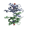 6ph2C 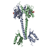 6ph3C  6ppsC 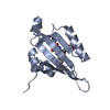 3t50S 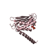 5epvS S: Starting model for refinement C: citing same article ( |
|---|---|
| Similar structure data |


-Links
 PDBj
PDBj


- Assembly
| Deposited unit | 
| ||||||||
|---|---|---|---|---|---|---|---|---|---|
| 1 |
| ||||||||
| Unit cell |
|
-Components
| #1: Protein | Mass: 54332.074 Da / Num. of mol.: 2 Source method: isolated from a genetically manipulated source Source: (gene. exp.) Brucella melitensis biotype 1 (strain 16M / ATCC 23456 / NCTC 10094) (bacteria)Strain: 16M / ATCC 23456 / NCTC 10094 / Gene: BMEII0679 / Plasmid: pET-24a / Production host: #2: Chemical |   Mass: 35.453 Da / Num. of mol.: 3 / Source method: obtained synthetically / Formula: Cl Mass: 35.453 Da / Num. of mol.: 3 / Source method: obtained synthetically / Formula: Cl#3: Chemical |   Mass: 456.344 Da / Num. of mol.: 2 / Source method: obtained synthetically / Formula: C17H21N4O9P / Feature type: SUBJECT OF INVESTIGATION Mass: 456.344 Da / Num. of mol.: 2 / Source method: obtained synthetically / Formula: C17H21N4O9P / Feature type: SUBJECT OF INVESTIGATION#4: Chemical | ChemComp-MG / |   Mass: 24.305 Da / Num. of mol.: 1 / Source method: obtained synthetically / Formula: Mg / Feature type: SUBJECT OF INVESTIGATION Mass: 24.305 Da / Num. of mol.: 1 / Source method: obtained synthetically / Formula: Mg / Feature type: SUBJECT OF INVESTIGATION#5: Chemical | ChemComp-ACP / |   Mass: 505.208 Da / Num. of mol.: 1 / Source method: obtained synthetically / Formula: C11H18N5O12P3 / Feature type: SUBJECT OF INVESTIGATION / Comment: AMP-PCP, energy-carrying molecule analogue*YM Mass: 505.208 Da / Num. of mol.: 1 / Source method: obtained synthetically / Formula: C11H18N5O12P3 / Feature type: SUBJECT OF INVESTIGATION / Comment: AMP-PCP, energy-carrying molecule analogue*YMHas ligand of interest | Y | |
|---|
-Experimental details
-Experiment
| Experiment | Method: X-RAY DIFFRACTION / Number of used crystals: 1 |
|---|
- Sample preparation
| Crystal | Density Matthews: 3.81 Å3/Da / Density % sol: 67.7 % |
|---|---|
| Crystal grow | Temperature: 294 K / Method: vapor diffusion, hanging drop / pH: 7.2 Details: 15% (v/v) MPD + 4% (w/v) PEG 4000 + 0.1 M Hepes, pH 7.2 |
-Data collection
| Diffraction | Mean temperature: 100 K / Serial crystal experiment: N |
|---|---|
| Diffraction source | Source: SYNCHROTRON / Site: SOLEIL  / Beamline: PROXIMA 2 / Wavelength: 0.9801 Å / Beamline: PROXIMA 2 / Wavelength: 0.9801 Å |
| Detector | Type: DECTRIS EIGER X 9M / Detector: PIXEL / Date: Nov 26, 2017 Details: CONVEX PREFOCUSSING MIRROR AND A KIRKPATRICK-BAEZ PAIR OF FOCUSSING MIRRORS |
| Radiation | Monochromator: CRYOGENICALLY COOLED CHANNEL CUT SI[111] CRYSTAL MONOCHROMATOR Protocol: SINGLE WAVELENGTH / Monochromatic (M) / Laue (L): M / Scattering type: x-ray |
| Radiation wavelength | Wavelength: 0.9801 Å / Relative weight: 1 |
| Reflection | Resolution: 3.25→62.53 Å / Num. obs: 26861 / % possible obs: 100 % / Redundancy: 14.8 % / Biso Wilson estimate: 92 Å2 / CC1/2: 0.998 / Rrim(I) all: 0.118 / Net I/σ(I): 13.1 |
| Reflection shell | Resolution: 3.25→3.47 Å / Redundancy: 15.3 % / Mean I/σ(I) obs: 1 / Num. unique obs: 4778 / CC1/2: 0.609 / Rrim(I) all: 2.774 / % possible all: 100 |
- Processing
| Software |
| |||||||||||||||||||||||||||||||||||||||||||||||||||||||||||||||||||||||||||||||||||||||||||||||||||||||||||||||||||||||||||||||||||||
|---|---|---|---|---|---|---|---|---|---|---|---|---|---|---|---|---|---|---|---|---|---|---|---|---|---|---|---|---|---|---|---|---|---|---|---|---|---|---|---|---|---|---|---|---|---|---|---|---|---|---|---|---|---|---|---|---|---|---|---|---|---|---|---|---|---|---|---|---|---|---|---|---|---|---|---|---|---|---|---|---|---|---|---|---|---|---|---|---|---|---|---|---|---|---|---|---|---|---|---|---|---|---|---|---|---|---|---|---|---|---|---|---|---|---|---|---|---|---|---|---|---|---|---|---|---|---|---|---|---|---|---|---|---|---|
| Refinement | Method to determine structure: MOLECULAR REPLACEMENT Starting model: 3T50 and 5EPV Resolution: 3.25→62.52 Å / SU ML: 0.6 / Cross valid method: THROUGHOUT / σ(F): 0.47 / Phase error: 38.45
| |||||||||||||||||||||||||||||||||||||||||||||||||||||||||||||||||||||||||||||||||||||||||||||||||||||||||||||||||||||||||||||||||||||
| Solvent computation | Shrinkage radii: 0.9 Å / VDW probe radii: 1.11 Å | |||||||||||||||||||||||||||||||||||||||||||||||||||||||||||||||||||||||||||||||||||||||||||||||||||||||||||||||||||||||||||||||||||||
| Refinement step | Cycle: LAST / Resolution: 3.25→62.52 Å
| |||||||||||||||||||||||||||||||||||||||||||||||||||||||||||||||||||||||||||||||||||||||||||||||||||||||||||||||||||||||||||||||||||||
| Refine LS restraints |
| |||||||||||||||||||||||||||||||||||||||||||||||||||||||||||||||||||||||||||||||||||||||||||||||||||||||||||||||||||||||||||||||||||||
| LS refinement shell |
|
