 Movie
Movie Controller

[English] 日本語
 Yorodumi
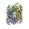
- PDB-6pd2: PntC-AEPT: fusion protein of phosphonate-specific cytidylyltransf... -
+ Open data

- Basic information
| Entry | Database: PDB / ID: 6pd2 | ||||||
|---|---|---|---|---|---|---|---|
| Title | PntC-AEPT: fusion protein of phosphonate-specific cytidylyltransferase and 2-aminoethylphosphonate (AEP) transaminase from Treponema denticola in complex with cytidine monophosphate-AEP | ||||||
 Components Components | Nucleotidyl transferase/aminotransferase, class V | ||||||
 Keywords Keywords | BIOSYNTHETIC PROTEIN / Phosphonate / Cytidylyltransferase / Cytidine monophosphate- 2-aminoethylphosphonate (CMP-AEP) | ||||||
| Function / homology |  Function and homology information Function and homology information(2-aminoethyl)phosphonate cytidylyltransferase / 2-aminoethylphosphonate-pyruvate transaminase / 2-aminoethylphosphonate:pyruvate transaminase activity / organic phosphonate biosynthetic process / organic phosphonate catabolic process / nucleotidyltransferase activity / metal ion binding Similarity search - Function | ||||||
| Biological species |  Treponema denticola (bacteria) Treponema denticola (bacteria) | ||||||
| Method |  X-RAY DIFFRACTION / SYNCHROTRON / MOLECULAR REPLACEMENT / Resolution: 1.95 Å X-RAY DIFFRACTION / SYNCHROTRON / MOLECULAR REPLACEMENT / Resolution: 1.95 Å | ||||||
 Authors Authors | Suits, M.D.L. / Whiteside, J. | ||||||
| Funding support |  Canada, 1items Canada, 1items
| ||||||
 Citation Citation | Journal: Nat Commun / Year: 2019 Title: The predominance of nucleotidyl activation in bacterial phosphonate biosynthesis. Authors: Rice, K. / Batul, K. / Whiteside, J. / Kelso, J. / Papinski, M. / Schmidt, E. / Pratasouskaya, A. / Wang, D. / Sullivan, R. / Bartlett, C. / Weadge, J.T. / Van der Kamp, M.W. / Moreno- ...Authors: Rice, K. / Batul, K. / Whiteside, J. / Kelso, J. / Papinski, M. / Schmidt, E. / Pratasouskaya, A. / Wang, D. / Sullivan, R. / Bartlett, C. / Weadge, J.T. / Van der Kamp, M.W. / Moreno-Hagelsieb, G. / Suits, M.D. / Horsman, G.P. | ||||||
| History |
|
- Structure visualization
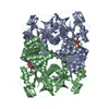
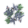
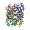
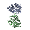
| Structure viewer | Molecule: MolmilJmol/JSmol |
|---|
- Downloads & links
-Download
| PDBx/mmCIF format | 6pd2.cif.gz | 528.3 KB | Display | PDBx/mmCIF format |
|---|---|---|---|---|
| PDB format | pdb6pd2.ent.gz | 425.4 KB | Display | PDB format |
| PDBx/mmJSON format | 6pd2.json.gz | Tree view | PDBx/mmJSON format | |
| Others |  Other downloads Other downloads |
-Validation report
| Arichive directory | https://data.pdbj.org/pub/pdb/validation_reports/pd/6pd2ftp://data.pdbj.org/pub/pdb/validation_reports/pd/6pd2 | HTTPS FTP |
|---|
-Related structure data
| Related structure data |  6pd1C  1jykS  1vjoS S: Starting model for refinement C: citing same article ( |
|---|---|
| Similar structure data |


-Links
 PDBj
PDBj

- Assembly
| Deposited unit | 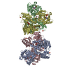
| ||||||||
|---|---|---|---|---|---|---|---|---|---|
| 1 | 
| ||||||||
| 2 | 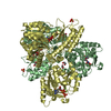
| ||||||||
| Unit cell |
|
-Components
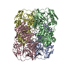
-Protein , 1 types, 4 molecules ABCD
| #1: Protein | Mass: 70148.680 Da / Num. of mol.: 4 Source method: isolated from a genetically manipulated source Source: (gene. exp.) Treponema denticola (strain ATCC 35405 / CIP 103919 / DSM 14222) (bacteria)Strain: ATCC 35405 / CIP 103919 / DSM 14222 / Gene: TDE_1415 / Production host: |
|---|
-Non-polymers , 8 types, 1506 molecules 


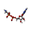


| #2: Chemical | ChemComp-PLP /  Mass: 247.142 Da / Num. of mol.: 4 / Source method: obtained synthetically / Formula: C8H10NO6P Mass: 247.142 Da / Num. of mol.: 4 / Source method: obtained synthetically / Formula: C8H10NO6P#3: Chemical | ChemComp-MG /  Mass: 24.305 Da / Num. of mol.: 8 / Source method: obtained synthetically / Formula: Mg Mass: 24.305 Da / Num. of mol.: 8 / Source method: obtained synthetically / Formula: Mg#4: Chemical | ChemComp-PO4 /  Mass: 94.971 Da / Num. of mol.: 11 / Source method: obtained synthetically / Formula: PO4 Mass: 94.971 Da / Num. of mol.: 11 / Source method: obtained synthetically / Formula: PO4#5: Chemical | ChemComp-0RC /  Mass: 430.245 Da / Num. of mol.: 4 / Source method: obtained synthetically / Formula: C11H20N4O10P2 / Feature type: SUBJECT OF INVESTIGATION Mass: 430.245 Da / Num. of mol.: 4 / Source method: obtained synthetically / Formula: C11H20N4O10P2 / Feature type: SUBJECT OF INVESTIGATION#6: Chemical | ChemComp-EDO /  Mass: 62.068 Da / Num. of mol.: 27 / Source method: obtained synthetically / Formula: C2H6O2 Mass: 62.068 Da / Num. of mol.: 27 / Source method: obtained synthetically / Formula: C2H6O2#7: Chemical |  Mass: 59.044 Da / Num. of mol.: 3 / Source method: obtained synthetically / Formula: C2H3O2 Mass: 59.044 Da / Num. of mol.: 3 / Source method: obtained synthetically / Formula: C2H3O2#8: Chemical |  Mass: 92.094 Da / Num. of mol.: 2 / Source method: obtained synthetically / Formula: C3H8O3 Mass: 92.094 Da / Num. of mol.: 2 / Source method: obtained synthetically / Formula: C3H8O3#9: Water | ChemComp-HOH / | Mass: 18.015 Da / Num. of mol.: 1447 / Source method: isolated from a natural source / Formula: H2O |
|---|
-Details
| Has ligand of interest | Y |
|---|
-Experimental details
-Experiment
| Experiment | Method: X-RAY DIFFRACTION / Number of used crystals: 1 |
|---|
- Sample preparation
| Crystal | Density Matthews: 2.82 Å3/Da / Density % sol: 56.44 % |
|---|---|
| Crystal grow | Temperature: 293 K / Method: vapor diffusion, hanging drop / pH: 6.5 Details: 0.2 M magnesium acetate tetrahydrate, 0.1 M sodium cacodylate trihydrate pH 6.5, 20% (w/v) polyethylene glycol 8,000 |
-Data collection
| Diffraction | Mean temperature: 100 K / Serial crystal experiment: N |
|---|---|
| Diffraction source | Source: SYNCHROTRON / Site: CLSI / Beamline: 08ID-1 / Wavelength: 0.97949 Å |
| Detector | Type: DECTRIS PILATUS3 S 6M / Detector: PIXEL / Date: Jun 10, 2017 |
| Radiation | Protocol: SINGLE WAVELENGTH / Monochromatic (M) / Laue (L): M / Scattering type: x-ray |
| Radiation wavelength | Wavelength: 0.97949 Å / Relative weight: 1 |
| Reflection | Resolution: 1.95→48 Å / Num. obs: 221625 / % possible obs: 98.2 % / Redundancy: 6.8 % / CC1/2: 0.989 / Net I/σ(I): 9.8 |
| Reflection shell | Resolution: 1.95→2.06 Å / Num. unique obs: 31932 |
- Processing
| Software |
| |||||||||||||||||||||||||||||||||||||||||||||||||||||||||||||||||||||||||||||||||||||||||||||||||||||||||||||||||||||||||||||||||||||||||||||||||||||||||||||||||||||||||||||||||||||||||||||||||||||||||||||||||||||||||
|---|---|---|---|---|---|---|---|---|---|---|---|---|---|---|---|---|---|---|---|---|---|---|---|---|---|---|---|---|---|---|---|---|---|---|---|---|---|---|---|---|---|---|---|---|---|---|---|---|---|---|---|---|---|---|---|---|---|---|---|---|---|---|---|---|---|---|---|---|---|---|---|---|---|---|---|---|---|---|---|---|---|---|---|---|---|---|---|---|---|---|---|---|---|---|---|---|---|---|---|---|---|---|---|---|---|---|---|---|---|---|---|---|---|---|---|---|---|---|---|---|---|---|---|---|---|---|---|---|---|---|---|---|---|---|---|---|---|---|---|---|---|---|---|---|---|---|---|---|---|---|---|---|---|---|---|---|---|---|---|---|---|---|---|---|---|---|---|---|---|---|---|---|---|---|---|---|---|---|---|---|---|---|---|---|---|---|---|---|---|---|---|---|---|---|---|---|---|---|---|---|---|---|---|---|---|---|---|---|---|---|---|---|---|---|---|---|---|---|
| Refinement | Method to determine structure: MOLECULAR REPLACEMENT Starting model: 1JYK, 1VJO Resolution: 1.95→47.976 Å / SU ML: 0.26 / Cross valid method: FREE R-VALUE / σ(F): 1.35 / Phase error: 29.23
| |||||||||||||||||||||||||||||||||||||||||||||||||||||||||||||||||||||||||||||||||||||||||||||||||||||||||||||||||||||||||||||||||||||||||||||||||||||||||||||||||||||||||||||||||||||||||||||||||||||||||||||||||||||||||
| Solvent computation | Shrinkage radii: 0.9 Å / VDW probe radii: 1.11 Å | |||||||||||||||||||||||||||||||||||||||||||||||||||||||||||||||||||||||||||||||||||||||||||||||||||||||||||||||||||||||||||||||||||||||||||||||||||||||||||||||||||||||||||||||||||||||||||||||||||||||||||||||||||||||||
| Refinement step | Cycle: LAST / Resolution: 1.95→47.976 Å
| |||||||||||||||||||||||||||||||||||||||||||||||||||||||||||||||||||||||||||||||||||||||||||||||||||||||||||||||||||||||||||||||||||||||||||||||||||||||||||||||||||||||||||||||||||||||||||||||||||||||||||||||||||||||||
| Refine LS restraints |
| |||||||||||||||||||||||||||||||||||||||||||||||||||||||||||||||||||||||||||||||||||||||||||||||||||||||||||||||||||||||||||||||||||||||||||||||||||||||||||||||||||||||||||||||||||||||||||||||||||||||||||||||||||||||||
| LS refinement shell |
|
