 Movie
Movie Controller

[English] 日本語
 Yorodumi
- PDB-6pc1: Crystal structure of Helicobacter pylori PPX/GppA (E143A) in comp... -
+ Open data

- Basic information
| Entry | Database: PDB / ID: 6pc1 | ||||||
|---|---|---|---|---|---|---|---|
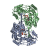
| Title | Crystal structure of Helicobacter pylori PPX/GppA (E143A) in complex with ppGpp | ||||||
 Components Components | Guanosine pentaphosphate phosphohydrolase | ||||||
 Keywords Keywords | HYDROLASE / pppGpp / ppGpp / PPX / GppA / Polyphosphate | ||||||
| Function / homology |  Function and homology information Function and homology information | ||||||
| Biological species |   Helicobacter pylori (bacteria) Helicobacter pylori (bacteria) | ||||||
| Method |  X-RAY DIFFRACTION / SYNCHROTRON / MOLECULAR REPLACEMENT / Resolution: 2.76 Å X-RAY DIFFRACTION / SYNCHROTRON / MOLECULAR REPLACEMENT / Resolution: 2.76 Å | ||||||
 Authors Authors | Song, H. / Wang, C. / Shaw, G.X. / Ji, X. | ||||||
 Citation Citation | Journal: Febs J. / Year: 2020 Title: Structure and activity of PPX/GppA homologs from Escherichia coli and Helicobacter pylori. Authors: Song, H. / Dharmasena, M.N. / Wang, C. / Shaw, G.X. / Cherry, S. / Tropea, J.E. / Jin, D.J. / Ji, X. | ||||||
| History |
|
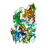
- Structure visualization
| Structure viewer | Molecule: MolmilJmol/JSmol |
|---|
- Downloads & links
-Download
| PDBx/mmCIF format | 6pc1.cif.gz | 394.5 KB | Display | PDBx/mmCIF format |
|---|---|---|---|---|
| PDB format | pdb6pc1.ent.gz | 318.3 KB | Display | PDB format |
| PDBx/mmJSON format | 6pc1.json.gz | Tree view | PDBx/mmJSON format | |
| Others |  Other downloads Other downloads |
-Validation report
| Arichive directory | https://data.pdbj.org/pub/pdb/validation_reports/pc/6pc1ftp://data.pdbj.org/pub/pdb/validation_reports/pc/6pc1 | HTTPS FTP |
|---|
-Related structure data
| Related structure data | 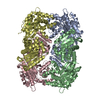 6pbzC 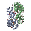 6pc0C 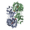 6pc2C 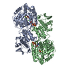 6pc3C 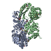 3hi0S S: Starting model for refinement C: citing same article ( |
|---|---|
| Similar structure data |


-Links
 PDBj
PDBj
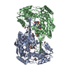
- Assembly
| Deposited unit | 
| ||||||||
|---|---|---|---|---|---|---|---|---|---|
| 1 | 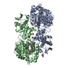
| ||||||||
| 2 | 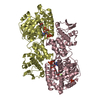
| ||||||||
| Unit cell |
|
-Components
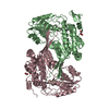
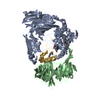
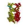
-Protein , 1 types, 4 molecules ABCD
| #1: Protein | Mass: 55597.512 Da / Num. of mol.: 4 / Mutation: E143A Source method: isolated from a genetically manipulated source Source: (gene. exp.) Helicobacter pylori (strain G27) (bacteria)Strain: G27 / Gene: HPG27_257 / Production host: |
|---|
-Non-polymers , 6 types, 388 molecules 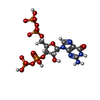


| #2: Chemical | ChemComp-G4P /  Type: RNA linking / Mass: 603.160 Da / Num. of mol.: 4 / Source method: obtained synthetically / Formula: C10H17N5O17P4 / Feature type: SUBJECT OF INVESTIGATION Type: RNA linking / Mass: 603.160 Da / Num. of mol.: 4 / Source method: obtained synthetically / Formula: C10H17N5O17P4 / Feature type: SUBJECT OF INVESTIGATION#3: Chemical | ChemComp-PO4 /  Mass: 94.971 Da / Num. of mol.: 5 / Source method: obtained synthetically / Formula: PO4 Mass: 94.971 Da / Num. of mol.: 5 / Source method: obtained synthetically / Formula: PO4#4: Chemical | ChemComp-LMR / (  Mass: 134.087 Da / Num. of mol.: 5 / Source method: obtained synthetically / Formula: C4H6O5 Mass: 134.087 Da / Num. of mol.: 5 / Source method: obtained synthetically / Formula: C4H6O5#5: Chemical |  Mass: 62.068 Da / Num. of mol.: 2 / Source method: obtained synthetically / Formula: C2H6O2 Mass: 62.068 Da / Num. of mol.: 2 / Source method: obtained synthetically / Formula: C2H6O2#6: Chemical |  Mass: 24.305 Da / Num. of mol.: 3 / Source method: obtained synthetically / Formula: Mg Mass: 24.305 Da / Num. of mol.: 3 / Source method: obtained synthetically / Formula: Mg#7: Water | ChemComp-HOH / | Mass: 18.015 Da / Num. of mol.: 369 / Source method: isolated from a natural source / Formula: H2O |
|---|
-Details
| Has ligand of interest | Y |
|---|
-Experimental details
-Experiment
| Experiment | Method: X-RAY DIFFRACTION / Number of used crystals: 1 |
|---|
- Sample preparation
| Crystal | Density Matthews: 2.91 Å3/Da / Density % sol: 57.69 % |
|---|---|
| Crystal grow | Temperature: 293 K / Method: vapor diffusion, hanging drop Details: 20% (v/v) PEG3350, 0.2 M Ammonium tartrate, 15 mM Malic acid |
-Data collection
| Diffraction | Mean temperature: 100 K / Serial crystal experiment: N |
|---|---|
| Diffraction source | Source: SYNCHROTRON / Site: APS  / Beamline: 22-ID / Wavelength: 1 Å / Beamline: 22-ID / Wavelength: 1 Å |
| Detector | Type: RAYONIX MX-300 / Detector: CCD / Date: Jun 6, 2014 |
| Radiation | Monochromator: Mirror1 / Protocol: SINGLE WAVELENGTH / Monochromatic (M) / Laue (L): M / Scattering type: x-ray |
| Radiation wavelength | Wavelength: 1 Å / Relative weight: 1 |
| Reflection | Resolution: 2.76→40 Å / Num. obs: 63095 / % possible obs: 99.3 % / Redundancy: 4.2 % / Rpim(I) all: 0.085 / Net I/σ(I): 7.4 |
| Reflection shell | Resolution: 2.7→2.8 Å / Redundancy: 1.9 % / Mean I/σ(I) obs: 0.7 / Num. unique obs: 5219 / % possible all: 74.6 |
- Processing
| Software |
| ||||||||||||||||||||||||||||||||||||||||||||||||||||||||
|---|---|---|---|---|---|---|---|---|---|---|---|---|---|---|---|---|---|---|---|---|---|---|---|---|---|---|---|---|---|---|---|---|---|---|---|---|---|---|---|---|---|---|---|---|---|---|---|---|---|---|---|---|---|---|---|---|---|
| Refinement | Method to determine structure: MOLECULAR REPLACEMENT Starting model: 3hi0 Resolution: 2.76→40 Å / SU ML: 0.37 / Cross valid method: FREE R-VALUE / σ(F): 1.35 / Phase error: 24.71
| ||||||||||||||||||||||||||||||||||||||||||||||||||||||||
| Solvent computation | Shrinkage radii: 0.9 Å / VDW probe radii: 1.11 Å | ||||||||||||||||||||||||||||||||||||||||||||||||||||||||
| Refinement step | Cycle: LAST / Resolution: 2.76→40 Å
| ||||||||||||||||||||||||||||||||||||||||||||||||||||||||
| Refine LS restraints |
| ||||||||||||||||||||||||||||||||||||||||||||||||||||||||
| LS refinement shell |
|
