 Movie
Movie Controller


+ Open data

- Basic information
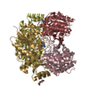
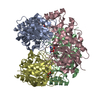
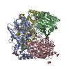
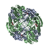
| Entry | Database: PDB / ID: 6pae | ||||||
|---|---|---|---|---|---|---|---|
| Title | Dickeya chrysanthemi complex with L-Asp at pH 5.6 | ||||||
 Components Components | L-asparaginase | ||||||
 Keywords Keywords | HYDROLASE / inactive mutant / hydrolysis of L-asparagine | ||||||
| Function / homology |  Function and homology information Function and homology information | ||||||
| Biological species |  Dickeya chrysanthemi (bacteria) Dickeya chrysanthemi (bacteria) | ||||||
| Method |  X-RAY DIFFRACTION / MOLECULAR REPLACEMENT / molecular replacement / Resolution: 1.6 Å X-RAY DIFFRACTION / MOLECULAR REPLACEMENT / molecular replacement / Resolution: 1.6 Å | ||||||
 Authors Authors | Lubkowski, J. / Wlodawer, A. | ||||||
 Citation Citation | Journal: Protein Sci. / Year: 2019 Title: Geometric considerations support the double-displacement catalytic mechanism of l-asparaginase. Authors: Lubkowski, J. / Wlodawer, A. | ||||||
| History |
|
- Structure visualization
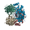
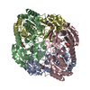
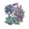
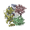
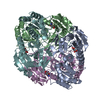
| Structure viewer | Molecule: MolmilJmol/JSmol |
|---|
- Downloads & links
-Download
| PDBx/mmCIF format | 6pae.cif.gz | 294.4 KB | Display | PDBx/mmCIF format |
|---|---|---|---|---|
| PDB format | pdb6pae.ent.gz | 239.2 KB | Display | PDB format |
| PDBx/mmJSON format | 6pae.json.gz | Tree view | PDBx/mmJSON format | |
| Others |  Other downloads Other downloads |
-Validation report
| Arichive directory | https://data.pdbj.org/pub/pdb/validation_reports/pa/6paeftp://data.pdbj.org/pub/pdb/validation_reports/pa/6pae | HTTPS FTP |
|---|
-Related structure data
| Related structure data |  6pa2C  6pa3C  6pa4C  6pa5C  6pa6C  6pa8C  6pa9C  6paaC  6pabC  6pacC C: citing same article ( |
|---|---|
| Similar structure data |

-Links
 PDBj
PDBj
- Assembly
| Deposited unit | 
| ||||||||
|---|---|---|---|---|---|---|---|---|---|
| 1 |
| ||||||||
| Unit cell |
|
-Components
| #1: Protein | Mass: 35123.020 Da / Num. of mol.: 4 Source method: isolated from a genetically manipulated source Details: Miller et al., FEBS Letters, 1993, 328, 275-279 / Source: (gene. exp.) Dickeya chrysanthemi (bacteria) / Gene: ansB, asn / Production host: #2: Chemical | ChemComp-ASP /   Type: L-peptide linking / Mass: 133.103 Da / Num. of mol.: 4 / Source method: obtained synthetically / Formula: C4H7NO4 / Feature type: SUBJECT OF INVESTIGATION Type: L-peptide linking / Mass: 133.103 Da / Num. of mol.: 4 / Source method: obtained synthetically / Formula: C4H7NO4 / Feature type: SUBJECT OF INVESTIGATION#3: Chemical | ChemComp-EDO /   Mass: 62.068 Da / Num. of mol.: 5 / Source method: obtained synthetically / Formula: C2H6O2 Mass: 62.068 Da / Num. of mol.: 5 / Source method: obtained synthetically / Formula: C2H6O2#4: Chemical | ChemComp-GOL / |   Mass: 92.094 Da / Num. of mol.: 1 / Source method: obtained synthetically / Formula: C3H8O3 Mass: 92.094 Da / Num. of mol.: 1 / Source method: obtained synthetically / Formula: C3H8O3#5: Water | ChemComp-HOH / |  Mass: 18.015 Da / Num. of mol.: 1384 / Source method: isolated from a natural source / Formula: H2O Mass: 18.015 Da / Num. of mol.: 1384 / Source method: isolated from a natural source / Formula: H2OHas ligand of interest | Y | |
|---|
-Experimental details
-Experiment
| Experiment | Method: X-RAY DIFFRACTION / Number of used crystals: 1 |
|---|
- Sample preparation
| Crystal | Density Matthews: 2.17 Å3/Da / Density % sol: 43.42 % |
|---|---|
| Crystal grow | Temperature: 298 K / Method: vapor diffusion, hanging drop / pH: 5.6 Details: see Miller et al., FEBS Letters, 1993, 328, 275-279 |
-Data collection
| Diffraction | Mean temperature: 100 K / Serial crystal experiment: N | ||||||||||||||||||||||||||||||||||||||||||||||||||||||||||||||||||
|---|---|---|---|---|---|---|---|---|---|---|---|---|---|---|---|---|---|---|---|---|---|---|---|---|---|---|---|---|---|---|---|---|---|---|---|---|---|---|---|---|---|---|---|---|---|---|---|---|---|---|---|---|---|---|---|---|---|---|---|---|---|---|---|---|---|---|---|
| Diffraction source | Source: ROTATING ANODE / Type: RIGAKU MICROMAX-007 HF / Wavelength: 1.5418 Å | ||||||||||||||||||||||||||||||||||||||||||||||||||||||||||||||||||
| Detector | Type: DECTRIS EIGER R 4M / Detector: PIXEL / Date: Sep 22, 2017 / Details: Multilayer X-ray mirrors VariMax HF | ||||||||||||||||||||||||||||||||||||||||||||||||||||||||||||||||||
| Radiation | Monochromator: Multilayer X-ray mirrors VariMax HF / Protocol: SINGLE WAVELENGTH / Monochromatic (M) / Laue (L): M / Scattering type: x-ray | ||||||||||||||||||||||||||||||||||||||||||||||||||||||||||||||||||
| Radiation wavelength | Wavelength: 1.5418 Å / Relative weight: 1 | ||||||||||||||||||||||||||||||||||||||||||||||||||||||||||||||||||
| Reflection | Resolution: 1.599→30 Å / Num. obs: 136750 / % possible obs: 85.8 % / Redundancy: 2.92 % / Rmerge(I) obs: 0.055 / Χ2: 1.207 / Net I/σ(I): 15.1 | ||||||||||||||||||||||||||||||||||||||||||||||||||||||||||||||||||
| Reflection shell |
|
-Phasing
| Phasing | Method: molecular replacement |
|---|
- Processing
| Software |
| |||||||||||||||||||||||||||||||||||||||||||||||||||||||||||||||||||||||||||
|---|---|---|---|---|---|---|---|---|---|---|---|---|---|---|---|---|---|---|---|---|---|---|---|---|---|---|---|---|---|---|---|---|---|---|---|---|---|---|---|---|---|---|---|---|---|---|---|---|---|---|---|---|---|---|---|---|---|---|---|---|---|---|---|---|---|---|---|---|---|---|---|---|---|---|---|---|
| Refinement | Method to determine structure: MOLECULAR REPLACEMENT / Resolution: 1.6→30 Å / Cor.coef. Fo:Fc: 0.976 / Cor.coef. Fo:Fc free: 0.964 / SU B: 1.523 / SU ML: 0.052 / SU R Cruickshank DPI: 0.084 / Cross valid method: THROUGHOUT / σ(F): 0 / ESU R: 0.084 / ESU R Free: 0.086 Details: HYDROGENS HAVE BEEN ADDED IN THE RIDING POSITIONS U VALUES : REFINED INDIVIDUALLY
| |||||||||||||||||||||||||||||||||||||||||||||||||||||||||||||||||||||||||||
| Solvent computation | Ion probe radii: 0.8 Å / Shrinkage radii: 0.8 Å / VDW probe radii: 1.2 Å | |||||||||||||||||||||||||||||||||||||||||||||||||||||||||||||||||||||||||||
| Displacement parameters | Biso max: 89.02 Å2 / Biso mean: 19.269 Å2 / Biso min: 6.87 Å2
| |||||||||||||||||||||||||||||||||||||||||||||||||||||||||||||||||||||||||||
| Refinement step | Cycle: final / Resolution: 1.6→30 Å
| |||||||||||||||||||||||||||||||||||||||||||||||||||||||||||||||||||||||||||
| Refine LS restraints |
| |||||||||||||||||||||||||||||||||||||||||||||||||||||||||||||||||||||||||||
| LS refinement shell | Resolution: 1.6→1.636 Å / Rfactor Rfree error: 0
|
