 Movie
Movie Controller

[English] 日本語
 Yorodumi
- PDB-1hfw: X-ray structure of the complex between Erwinia chrysanthemi L-asp... -
+ Open data

- Basic information
| Entry | Database: PDB / ID: 1hfw | ||||||
|---|---|---|---|---|---|---|---|
| Title | X-ray structure of the complex between Erwinia chrysanthemi L-asparaginase and L-Glutamate | ||||||
 Components Components | L-ASPARAGINASE | ||||||
 Keywords Keywords | ASPARAGINASE / HYDROLASE / COMPLEX / D-ASPARTATE | ||||||
| Function / homology |  Function and homology information Function and homology information | ||||||
| Biological species |  ERWINIA CHRYSANTHEMI (bacteria) ERWINIA CHRYSANTHEMI (bacteria) | ||||||
| Method |  X-RAY DIFFRACTION / MOLECULAR REPLACEMENT / Resolution: 1.8 Å X-RAY DIFFRACTION / MOLECULAR REPLACEMENT / Resolution: 1.8 Å | ||||||
 Authors Authors | Lubkowski, J. / Wlodawer, A. / Kolyani, K.A. | ||||||
 Citation Citation | Journal: Biochemistry / Year: 2001 Title: Stuctural Basis for the Activity and Substrate Specificity of Erwinia Chrysanthemi L-Asparaginase Authors: Kolyani, K.A. / Wlodawer, A. / Lubkowski, J. #1: Journal: FEBS Lett. / Year: 1993 Title: A Left-Handed Crossover Involved in Amidohydrolase Catalysis, Crystal Structure of Erwinia Chrysanthemi L-Asparaginase with Bound L-Aspartate Authors: Miller, M. / Rao, J.K.M. / Wlodawer, A. / Gribskov, M.R. #2: Journal: Proc.Natl.Acad.Sci.USA / Year: 1993Title: Crystal Structure of Escherichia Coli L-Asparaginase, an Enzyme Used in Cancer Therapy Authors: Swain, A.L. / Jaskolski, M. / Housset, D. / Rao, J.K.M. / Wlodawer, A. | ||||||
| History |
|
- Structure visualization
| Structure viewer | Molecule: MolmilJmol/JSmol |
|---|
- Downloads & links
-Download
| PDBx/mmCIF format | 1hfw.cif.gz | 255.4 KB | Display | PDBx/mmCIF format |
|---|---|---|---|---|
| PDB format | pdb1hfw.ent.gz | 208.1 KB | Display | PDB format |
| PDBx/mmJSON format | 1hfw.json.gz | Tree view | PDBx/mmJSON format | |
| Others |  Other downloads Other downloads |
-Validation report
| Arichive directory | https://data.pdbj.org/pub/pdb/validation_reports/hf/1hfwftp://data.pdbj.org/pub/pdb/validation_reports/hf/1hfw | HTTPS FTP |
|---|
-Related structure data


-Links
 PDBj
PDBj

- Assembly
| Deposited unit | 
| ||||||||
|---|---|---|---|---|---|---|---|---|---|
| 1 |
| ||||||||
| Unit cell |
| ||||||||
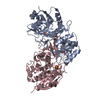
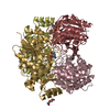
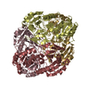
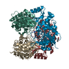
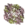
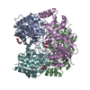
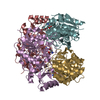
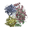
| Details | BIOLOGICAL_UNIT: HOMOTETRAMER |
-Components
| #1: Protein | Mass: 35123.020 Da / Num. of mol.: 4 / Source method: isolated from a natural source Details: THE NEW NAME OF ERWINIA CHRYSANTHEMI IS PECTOBACTERIUM CHRYSANTHEMI Source: (natural) ERWINIA CHRYSANTHEMI (bacteria) / Strain: NCPPB 1125 / References: UniProt: P06608, asparaginase#2: Chemical | ChemComp-GLU /   Type: L-peptide linking / Mass: 147.129 Da / Num. of mol.: 4 / Source method: obtained synthetically / Formula: C5H9NO4 Type: L-peptide linking / Mass: 147.129 Da / Num. of mol.: 4 / Source method: obtained synthetically / Formula: C5H9NO4#3: Water | ChemComp-HOH / |  Mass: 18.015 Da / Num. of mol.: 961 / Source method: isolated from a natural source / Formula: H2O Mass: 18.015 Da / Num. of mol.: 961 / Source method: isolated from a natural source / Formula: H2O |
|---|
-Experimental details
-Experiment
| Experiment | Method: X-RAY DIFFRACTION / Number of used crystals: 1 |
|---|
- Sample preparation
| Crystal | Density Matthews: 2.31 Å3/Da / Density % sol: 46.77 % | ||||||||||||||||||||
|---|---|---|---|---|---|---|---|---|---|---|---|---|---|---|---|---|---|---|---|---|---|
| Crystal grow | pH: 5.4 Details: 40%(W/V) AMMONIUM SULFATE, 2%(V/V) PEG400, 0.1M TRIS (PH 8.5), CROSSLINKING WITH 0.1% GLUTARALDEHYDE. TRANSFER TO AMMONIUM SULFATE-FREE, 30% PEG4000, CHANGE OF THE BUFFER TO 0.1M SODIUM ACETATE | ||||||||||||||||||||
| Crystal grow | *PLUS Method: vapor diffusion, hanging drop / Details: Miller, M., (1993) FEBS Lett., 328, 275. / PH range low: 9 / PH range high: 8 | ||||||||||||||||||||
| Components of the solutions | *PLUS
|
-Data collection
| Diffraction | Mean temperature: 295 K |
|---|---|
| Diffraction source | Source: ROTATING ANODE / Type: RIGAKU RU200 / Wavelength: 1.5478 |
| Detector | Type: MAR scanner 345 mm plate / Detector: IMAGE PLATE / Date: Aug 15, 1998 / Details: COLLIMATOR |
| Radiation | Monochromator: GRAPHITE CRYSTAL / Protocol: SINGLE WAVELENGTH / Monochromatic (M) / Laue (L): M / Scattering type: x-ray |
| Radiation wavelength | Wavelength: 1.5478 Å / Relative weight: 1 |
| Reflection | Resolution: 1.8→40 Å / Num. obs: 102023 / % possible obs: 86 % / Observed criterion σ(I): -3 / Redundancy: 2.8 % / Rsym value: 0.076 / Net I/σ(I): 7.4 |
| Reflection shell | Resolution: 1.8→1.86 Å / Mean I/σ(I) obs: 2.8 / Rsym value: 0.337 / % possible all: 65.4 |
| Reflection | *PLUS Num. measured all: 270156 / Rmerge(I) obs: 0.076 |
| Reflection shell | *PLUS % possible obs: 65.4 % / Rmerge(I) obs: 0.337 |
- Processing
| Software |
| ||||||||||||||||||||||||||||||||||||||||||||||||||||||||||||||||||||||||||||||||
|---|---|---|---|---|---|---|---|---|---|---|---|---|---|---|---|---|---|---|---|---|---|---|---|---|---|---|---|---|---|---|---|---|---|---|---|---|---|---|---|---|---|---|---|---|---|---|---|---|---|---|---|---|---|---|---|---|---|---|---|---|---|---|---|---|---|---|---|---|---|---|---|---|---|---|---|---|---|---|---|---|---|
| Refinement | Method to determine structure: MOLECULAR REPLACEMENT Starting model: PREVIUOSLY PUBLISHED STRUCTURE OF ERWINIA CHRYSANTHEMI L-ASPARAGINSE (MILLER ET AL., FEBS LETT., 1993 Resolution: 1.8→10 Å / Rfactor Rfree error: 0.0045 / Data cutoff high absF: 100000 / Isotropic thermal model: RESTRAINED / Cross valid method: THROUGHOUT / σ(F): 2
| ||||||||||||||||||||||||||||||||||||||||||||||||||||||||||||||||||||||||||||||||
| Solvent computation | Solvent model: DENSITY MODIFICATION / Bsol: 84.82 Å2 / ksol: 0.421 e/Å3 | ||||||||||||||||||||||||||||||||||||||||||||||||||||||||||||||||||||||||||||||||
| Displacement parameters | Biso mean: 19.4 Å2
| ||||||||||||||||||||||||||||||||||||||||||||||||||||||||||||||||||||||||||||||||
| Refine analyze |
| ||||||||||||||||||||||||||||||||||||||||||||||||||||||||||||||||||||||||||||||||
| Refinement step | Cycle: LAST / Resolution: 1.8→10 Å
| ||||||||||||||||||||||||||||||||||||||||||||||||||||||||||||||||||||||||||||||||
| Refine LS restraints |
| ||||||||||||||||||||||||||||||||||||||||||||||||||||||||||||||||||||||||||||||||
| LS refinement shell | Resolution: 1.8→1.86 Å / Rfactor Rfree error: 0.031 / Total num. of bins used: 10
| ||||||||||||||||||||||||||||||||||||||||||||||||||||||||||||||||||||||||||||||||
| Xplor file |
| ||||||||||||||||||||||||||||||||||||||||||||||||||||||||||||||||||||||||||||||||
| Software | *PLUS Name: CNS / Version: 1 / Classification: refinement | ||||||||||||||||||||||||||||||||||||||||||||||||||||||||||||||||||||||||||||||||
| Refine LS restraints | *PLUS
|
