 Movie
Movie Controller

[English] 日本語
 Yorodumi
- PDB-2hln: L-asparaginase from Erwinia carotovora in complex with glutamic acid -
+ Open data

- Basic information
| Entry | Database: PDB / ID: 2hln | ||||||
|---|---|---|---|---|---|---|---|
| Title | L-asparaginase from Erwinia carotovora in complex with glutamic acid | ||||||
 Components Components | L-asparaginase | ||||||
 Keywords Keywords | HYDROLASE / L-asparaginase / Erwinia carotovora | ||||||
| Function / homology |  Function and homology information Function and homology information | ||||||
| Biological species |  Pectobacterium atrosepticum (bacteria) Pectobacterium atrosepticum (bacteria) | ||||||
| Method |  X-RAY DIFFRACTION / SYNCHROTRON / MOLECULAR REPLACEMENT / Resolution: 2.2 Å X-RAY DIFFRACTION / SYNCHROTRON / MOLECULAR REPLACEMENT / Resolution: 2.2 Å | ||||||
 Authors Authors | Kravchenko, O.V. / Kislitsin, Y.A. / Popov, A.N. / Nikonov, S.V. / Kuranova, I.P. | ||||||
 Citation Citation | Journal: Acta Crystallogr.,Sect.D / Year: 2008 Title: Three-dimensional structures of L-asparaginase from Erwinia carotovora complexed with aspartate and glutamate. Authors: Kravchenko, O.V. / Kislitsin, Y.A. / Popov, A.N. / Nikonov, S.V. / Kuranova, I.P. | ||||||
| History |
|
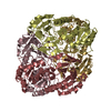
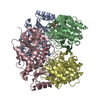
- Structure visualization
| Structure viewer | Molecule: MolmilJmol/JSmol |
|---|
- Downloads & links
-Download
| PDBx/mmCIF format | 2hln.cif.gz | 721 KB | Display | PDBx/mmCIF format |
|---|---|---|---|---|
| PDB format | pdb2hln.ent.gz | 595.6 KB | Display | PDB format |
| PDBx/mmJSON format | 2hln.json.gz | Tree view | PDBx/mmJSON format | |
| Others |  Other downloads Other downloads |
-Validation report
| Arichive directory | https://data.pdbj.org/pub/pdb/validation_reports/hl/2hlnftp://data.pdbj.org/pub/pdb/validation_reports/hl/2hln | HTTPS FTP |
|---|
-Related structure data
| Related structure data |  2gvnC  1zcfS S: Starting model for refinement C: citing same article ( |
|---|---|
| Similar structure data |


-Links
 PDBj
PDBj

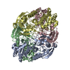
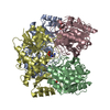
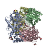
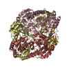
- Assembly
| Deposited unit | 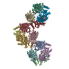
| ||||||||
|---|---|---|---|---|---|---|---|---|---|
| 1 | 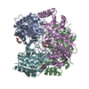
| ||||||||
| 2 | 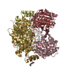
| ||||||||
| 3 | 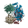
| ||||||||
| Unit cell |
| ||||||||
| Details | The biological unit is tetramer. There are 3 biological units in the asymmetric unit. The biological unit 1 consists of chains A,B,E,F. The biological unit 2 consists of chains C,D,G,H. The biological unit 3 consists of chains I,J,K,L. |
-Components
| #1: Protein | Mass: 34528.266 Da / Num. of mol.: 12 Source method: isolated from a genetically manipulated source Source: (gene. exp.) Pectobacterium atrosepticum (bacteria) / Gene: lanS / Plasmid: pACYCLANS / Species (production host): Escherichia coli / Production host: #2: Chemical | ChemComp-GLU /   Type: L-peptide linking / Mass: 147.129 Da / Num. of mol.: 12 / Source method: obtained synthetically / Formula: C5H9NO4 Type: L-peptide linking / Mass: 147.129 Da / Num. of mol.: 12 / Source method: obtained synthetically / Formula: C5H9NO4#3: Chemical | ChemComp-PEG /   Mass: 106.120 Da / Num. of mol.: 8 / Source method: obtained synthetically / Formula: C4H10O3 Mass: 106.120 Da / Num. of mol.: 8 / Source method: obtained synthetically / Formula: C4H10O3#4: Water | ChemComp-HOH / |  Mass: 18.015 Da / Num. of mol.: 1878 / Source method: isolated from a natural source / Formula: H2O Mass: 18.015 Da / Num. of mol.: 1878 / Source method: isolated from a natural source / Formula: H2O |
|---|
-Experimental details
-Experiment
| Experiment | Method: X-RAY DIFFRACTION / Number of used crystals: 1 |
|---|
- Sample preparation
| Crystal | Density Matthews: 2.39 Å3/Da / Density % sol: 48.51 % |
|---|---|
| Crystal grow | Temperature: 295 K / Method: vapor diffusion, hanging drop / pH: 6.5 Details: 0.1% sodium cacodilate, 8% PEG MME 5000, 0.06% octylglucoside, 0.08M glutamic acid, 0.02% sodium azide, pH 6.5, VAPOR DIFFUSION, HANGING DROP, temperature 295K |
-Data collection
| Diffraction | Mean temperature: 100 K |
|---|---|
| Diffraction source | Source: SYNCHROTRON / Site: EMBL/DESY, HAMBURG  / Beamline: BW7A / Wavelength: 0.99 / Beamline: BW7A / Wavelength: 0.99 |
| Detector | Type: MAR CCD 165 mm / Detector: CCD / Date: Oct 24, 2005 / Details: bent mirror |
| Radiation | Monochromator: double crystal focussing monochromator / Protocol: SINGLE WAVELENGTH / Monochromatic (M) / Laue (L): M / Scattering type: x-ray |
| Radiation wavelength | Wavelength: 0.99 Å / Relative weight: 1 |
| Reflection | Resolution: 2.2→20 Å / Num. all: 180862 / Num. obs: 180862 / % possible obs: 91.8 % / Observed criterion σ(F): 0 / Observed criterion σ(I): 3 / Redundancy: 3.3 % / Rmerge(I) obs: 0.08 / Net I/σ(I): 17.2 |
| Reflection shell | Resolution: 2.2→2.22 Å / Redundancy: 3.3 % / Rmerge(I) obs: 0.423 / Mean I/σ(I) obs: 2.6 / Num. unique all: 5910 / % possible all: 91 |
- Processing
| Software |
| ||||||||||||||||||||||||||||||||||||||||||||||||||||||||||||||||||||||||||||||||||||||||||
|---|---|---|---|---|---|---|---|---|---|---|---|---|---|---|---|---|---|---|---|---|---|---|---|---|---|---|---|---|---|---|---|---|---|---|---|---|---|---|---|---|---|---|---|---|---|---|---|---|---|---|---|---|---|---|---|---|---|---|---|---|---|---|---|---|---|---|---|---|---|---|---|---|---|---|---|---|---|---|---|---|---|---|---|---|---|---|---|---|---|---|---|
| Refinement | Method to determine structure: MOLECULAR REPLACEMENT Starting model: pdb entry 1ZCF Resolution: 2.2→19.98 Å / Cor.coef. Fo:Fc: 0.946 / Cor.coef. Fo:Fc free: 0.918 / SU B: 7.208 / SU ML: 0.18 / Cross valid method: THROUGHOUT / σ(F): 0 / σ(I): 0 / ESU R: 0.385 / ESU R Free: 0.258 / Stereochemistry target values: MAXIMUM LIKELIHOOD / Details: HYDROGENS HAVE BEEN ADDED IN THE RIDING POSITIONS
| ||||||||||||||||||||||||||||||||||||||||||||||||||||||||||||||||||||||||||||||||||||||||||
| Solvent computation | Ion probe radii: 0.8 Å / Shrinkage radii: 0.8 Å / VDW probe radii: 1.4 Å / Solvent model: MASK | ||||||||||||||||||||||||||||||||||||||||||||||||||||||||||||||||||||||||||||||||||||||||||
| Displacement parameters | Biso mean: 43.142 Å2
| ||||||||||||||||||||||||||||||||||||||||||||||||||||||||||||||||||||||||||||||||||||||||||
| Refine analyze |
| ||||||||||||||||||||||||||||||||||||||||||||||||||||||||||||||||||||||||||||||||||||||||||
| Refinement step | Cycle: LAST / Resolution: 2.2→19.98 Å
| ||||||||||||||||||||||||||||||||||||||||||||||||||||||||||||||||||||||||||||||||||||||||||
| Refine LS restraints |
| ||||||||||||||||||||||||||||||||||||||||||||||||||||||||||||||||||||||||||||||||||||||||||
| LS refinement shell | Resolution: 2.2→2.256 Å / Total num. of bins used: 20
|
