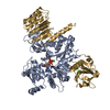
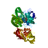
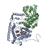
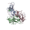
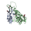
Entry Database : PDB / ID : 6p8iTitle N-terminal 5 domains of IGFIIR Cation-independent mannose-6-phosphate receptor Keywords / / / / Function / homology Function Domain/homology Component
/ / / / / / / / / / / / / / / / / / / / / / / / / / / / / / / / / / / / / / / / / / / / / / / / / / / / / / / / / / / / / / / / / / / / / / / / / / / / / / / / / / Biological species Homo sapiens (human)Method / / / Resolution : 2.54 Å Authors Olson, L.J. / Dahms, N.M. / Kim, J.-J.P. Funding support Organization Grant number Country National Institutes of Health/National Institute of Diabetes and Digestive and Kidney Disease (NIH/NIDDK) RO1DK042667
Journal : Commun Biol / Year : 2020Title : Allosteric regulation of lysosomal enzyme recognition by the cation-independent mannose 6-phosphate receptor.Authors : Olson, L.J. / Misra, S.K. / Ishihara, M. / Battaile, K.P. / Grant, O.C. / Sood, A. / Woods, R.J. / Kim, J.P. / Tiemeyer, M. / Ren, G. / Sharp, J.S. / Dahms, N.M. History Deposition Jun 7, 2019 Deposition site / Processing site Revision 1.0 Jun 24, 2020 Provider / Type Revision 2.0 Jul 29, 2020 Group Advisory / Atomic model ... Advisory / Atomic model / Data collection / Derived calculations / Structure summary Category atom_site / chem_comp ... atom_site / chem_comp / entity / pdbx_branch_scheme / pdbx_chem_comp_identifier / pdbx_entity_branch / pdbx_entity_branch_descriptor / pdbx_entity_branch_link / pdbx_entity_branch_list / pdbx_entity_nonpoly / pdbx_nonpoly_scheme / pdbx_struct_assembly_gen / pdbx_validate_close_contact / struct_asym / struct_conn / struct_site / struct_site_gen Item _atom_site.auth_asym_id / _atom_site.auth_seq_id ... _atom_site.auth_asym_id / _atom_site.auth_seq_id / _atom_site.label_asym_id / _atom_site.label_entity_id / _chem_comp.name / _pdbx_struct_assembly_gen.asym_id_list / _pdbx_validate_close_contact.auth_asym_id_1 / _pdbx_validate_close_contact.auth_asym_id_2 / _pdbx_validate_close_contact.auth_seq_id_1 / _pdbx_validate_close_contact.auth_seq_id_2 / _struct_conn.pdbx_role / _struct_conn.ptnr1_auth_asym_id / _struct_conn.ptnr1_auth_seq_id / _struct_conn.ptnr1_label_asym_id / _struct_conn.ptnr2_auth_asym_id / _struct_conn.ptnr2_auth_seq_id / _struct_conn.ptnr2_label_asym_id Description / Provider / Type Revision 2.1 Sep 30, 2020 Group / Derived calculations / Structure summaryCategory chem_comp / citation ... chem_comp / citation / citation_author / struct_conn Item _chem_comp.pdbx_synonyms / _citation.country ... _chem_comp.pdbx_synonyms / _citation.country / _citation.journal_abbrev / _citation.journal_id_CSD / _citation.journal_id_ISSN / _citation.journal_volume / _citation.page_first / _citation.page_last / _citation.pdbx_database_id_DOI / _citation.pdbx_database_id_PubMed / _citation.title / _citation.year / _struct_conn.pdbx_leaving_atom_flag Revision 2.2 Mar 16, 2022 Group / Database references / Category / pdbx_audit_supportItem / _database_2.pdbx_database_accession / _pdbx_audit_support.funding_organizationRevision 2.3 Oct 11, 2023 Group / Refinement descriptionCategory / chem_comp_bond / pdbx_initial_refinement_modelRevision 2.4 Nov 6, 2024 Group / Category / pdbx_modification_feature / Item
Show all Show less
 Movie
Movie Controller


 Open data
 Basic information
 Components
 Keywords
 Function and homology information
 Homo sapiens (human)
 X-RAY DIFFRACTION /
  Authors
 United States, 1items
  Citation
 Structure visualization
 Downloads & links
 Other downloads
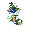


 PDBj
PDBj


 Assembly

 Spodoptera frugiperda (fall armyworm) / References: UniProt: P11717
 Mass: 18.015 Da / Num. of mol.: 28 / Source method: isolated from a natural source / Formula: H2O
 Sample preparation
 Processing