 Movie
Movie Controller

[English] 日本語
 Yorodumi
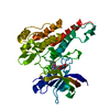
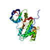
- PDB-6ozu: Crystal structure of the MIF4G domain of Trypanosoma cruzi transl... -
+ Open data

- Basic information
| Entry | Database: PDB / ID: 6ozu | ||||||
|---|---|---|---|---|---|---|---|
| Title | Crystal structure of the MIF4G domain of Trypanosoma cruzi translation initiation factor EIF4G5 | ||||||
 Components Components | Eukaryotic translation initiation factor 4 gamma 5 | ||||||
 Keywords Keywords | TRANSLATION / Initiation Factor / EIF4G | ||||||
| Function / homology | Initiation factor 4G / MIF4G domain / MIF4G-like, type 3 / translation initiation factor activity / Armadillo-type fold / RNA binding / Eukaryotic translation initiation factor 4 gamma 5 / MIF4G domain-containing protein Function and homology information Function and homology information | ||||||
| Biological species |  | ||||||
| Method |  X-RAY DIFFRACTION / SYNCHROTRON / SAD / Resolution: 2.4 Å X-RAY DIFFRACTION / SYNCHROTRON / SAD / Resolution: 2.4 Å | ||||||
 Authors Authors | Guimaraes, B.G. / Santos, L.P.C. | ||||||
 Citation Citation | Journal: Acta Crystallogr.,Sect.F / Year: 2019 Title: Crystal structure of the MIF4G domain of the Trypanosoma cruzi translation initiation factor EIF4G5. Authors: Camillo Dos Santos, L.P. / de Matos, B.M. / de Maman Ribeiro, B.C. / Zanchin, N.I.T. / Guimaraes, B.G. #1: Journal: Acta Crystallogr.,Sect.F / Year: 2019Title: Crystal structure of the MIF4G domain of the Trypanosoma cruzi translation initiation factor EIF4G5 Authors: Santos, L.P.C. / Matos, B.M. / Ribeiro, B.C.M. / Zanchin, N.I.T. / Guimaraes, B.G. | ||||||
| History |
|
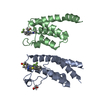
- Structure visualization
| Structure viewer | Molecule: MolmilJmol/JSmol |
|---|
- Downloads & links
-Download
| PDBx/mmCIF format | 6ozu.cif.gz | 212.2 KB | Display | PDBx/mmCIF format |
|---|---|---|---|---|
| PDB format | pdb6ozu.ent.gz | 172.2 KB | Display | PDB format |
| PDBx/mmJSON format | 6ozu.json.gz | Tree view | PDBx/mmJSON format | |
| Others |  Other downloads Other downloads |
-Validation report
| Summary document | 6ozu_validation.pdf.gz | 465 KB | Display | wwPDB validaton report |
|---|---|---|---|---|
| Full document | 6ozu_full_validation.pdf.gz | 470.3 KB | Display | |
| Data in XML | 6ozu_validation.xml.gz | 19.8 KB | Display | |
| Data in CIF | 6ozu_validation.cif.gz | 26.9 KB | Display | |
| Arichive directory | https://data.pdbj.org/pub/pdb/validation_reports/oz/6ozuftp://data.pdbj.org/pub/pdb/validation_reports/oz/6ozu | HTTPS FTP |
-Related structure data
| Similar structure data |
|---|


-Links
 PDBj
PDBj

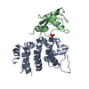
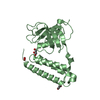
- Assembly
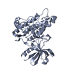
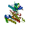
| Deposited unit | 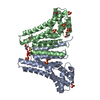
| ||||||||
|---|---|---|---|---|---|---|---|---|---|
| 1 | 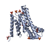
| ||||||||
| 2 | 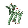
| ||||||||
| 3 |
| ||||||||
| Unit cell |
|
-Components
| #1: Protein | Mass: 28248.568 Da / Num. of mol.: 2 / Fragment: residues 142-371 Source method: isolated from a genetically manipulated source Source: (gene. exp.)  #2: Chemical | ChemComp-SO4 /   Mass: 96.063 Da / Num. of mol.: 13 / Source method: obtained synthetically / Formula: SO4 Mass: 96.063 Da / Num. of mol.: 13 / Source method: obtained synthetically / Formula: SO4#3: Chemical | ChemComp-GOL /   Mass: 92.094 Da / Num. of mol.: 6 / Source method: obtained synthetically / Formula: C3H8O3 Mass: 92.094 Da / Num. of mol.: 6 / Source method: obtained synthetically / Formula: C3H8O3#4: Water | ChemComp-HOH / |  Mass: 18.015 Da / Num. of mol.: 95 / Source method: isolated from a natural source / Formula: H2O Mass: 18.015 Da / Num. of mol.: 95 / Source method: isolated from a natural source / Formula: H2OHas ligand of interest | N | |
|---|
-Experimental details
-Experiment
| Experiment | Method: X-RAY DIFFRACTION / Number of used crystals: 1 |
|---|
- Sample preparation
| Crystal | Density Matthews: 3.3 Å3/Da / Density % sol: 62.72 % |
|---|---|
| Crystal grow | Temperature: 291.15 K / Method: vapor diffusion, sitting drop / pH: 6.5 / Details: sodium cacodylate 0.1 M, ammonium sulfate 2 M |
-Data collection
| Diffraction | Mean temperature: 100 K / Serial crystal experiment: N |
|---|---|
| Diffraction source | Source: SYNCHROTRON / Site: SOLEIL  / Beamline: PROXIMA 1 / Wavelength: 1.051 Å / Beamline: PROXIMA 1 / Wavelength: 1.051 Å |
| Detector | Type: DECTRIS PILATUS3 S 6M / Detector: PIXEL / Date: Jun 22, 2017 |
| Radiation | Protocol: SINGLE WAVELENGTH / Monochromatic (M) / Laue (L): M / Scattering type: x-ray |
| Radiation wavelength | Wavelength: 1.051 Å / Relative weight: 1 |
| Reflection | Resolution: 2.4→50 Å / Num. obs: 30916 / % possible obs: 99.7 % / Redundancy: 12.7 % / Biso Wilson estimate: 66.66 Å2 / CC1/2: 1 / Net I/σ(I): 19.66 |
| Reflection shell | Resolution: 2.4→2.54 Å / Redundancy: 13 % / Mean I/σ(I) obs: 0.94 / Num. unique obs: 62721 / CC1/2: 0.7 / % possible all: 98.4 |
- Processing
| Software |
| ||||||||||||||||||||||||||||||||||||||||||||||||||||||||||||||||||||||||||||||||||||||||||||||||||||||||||||
|---|---|---|---|---|---|---|---|---|---|---|---|---|---|---|---|---|---|---|---|---|---|---|---|---|---|---|---|---|---|---|---|---|---|---|---|---|---|---|---|---|---|---|---|---|---|---|---|---|---|---|---|---|---|---|---|---|---|---|---|---|---|---|---|---|---|---|---|---|---|---|---|---|---|---|---|---|---|---|---|---|---|---|---|---|---|---|---|---|---|---|---|---|---|---|---|---|---|---|---|---|---|---|---|---|---|---|---|---|---|
| Refinement | Method to determine structure: SAD / Resolution: 2.4→48.92 Å / Cor.coef. Fo:Fc: 0.931 / Cor.coef. Fo:Fc free: 0.897 / SU R Cruickshank DPI: 0.346 / Cross valid method: THROUGHOUT / σ(F): 0 / SU R Blow DPI: 0.37 / SU Rfree Blow DPI: 0.253 / SU Rfree Cruickshank DPI: 0.251
| ||||||||||||||||||||||||||||||||||||||||||||||||||||||||||||||||||||||||||||||||||||||||||||||||||||||||||||
| Displacement parameters | Biso max: 172.91 Å2 / Biso mean: 74.08 Å2 / Biso min: 35.06 Å2
| ||||||||||||||||||||||||||||||||||||||||||||||||||||||||||||||||||||||||||||||||||||||||||||||||||||||||||||
| Refine analyze | Luzzati coordinate error obs: 0.36 Å | ||||||||||||||||||||||||||||||||||||||||||||||||||||||||||||||||||||||||||||||||||||||||||||||||||||||||||||
| Refinement step | Cycle: final / Resolution: 2.4→48.92 Å
| ||||||||||||||||||||||||||||||||||||||||||||||||||||||||||||||||||||||||||||||||||||||||||||||||||||||||||||
| Refine LS restraints |
| ||||||||||||||||||||||||||||||||||||||||||||||||||||||||||||||||||||||||||||||||||||||||||||||||||||||||||||
| LS refinement shell | Resolution: 2.4→2.44 Å / Rfactor Rfree error: 0 / Total num. of bins used: 50
| ||||||||||||||||||||||||||||||||||||||||||||||||||||||||||||||||||||||||||||||||||||||||||||||||||||||||||||
| Refinement TLS params. | Method: refined / Refine-ID: X-RAY DIFFRACTION
| ||||||||||||||||||||||||||||||||||||||||||||||||||||||||||||||||||||||||||||||||||||||||||||||||||||||||||||
| Refinement TLS group |
|
