 Movie
Movie Controller


+ Open data

- Basic information
| Entry | Database: PDB / ID: 6oyz | ||||||
|---|---|---|---|---|---|---|---|
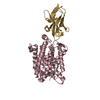
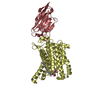
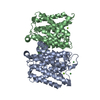
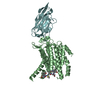
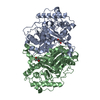
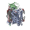
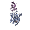
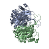
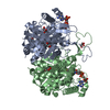
| Title | Crystal structure of MraY bound to capuramycin | ||||||
 Components Components |
| ||||||
 Keywords Keywords | MEMBRANE PROTEIN/TRANSFERASE / peptidoglycan biosynthesis / antibiotic target / integral membrane enzyme / translocase / MEMBRANE PROTEIN / MEMBRANE PROTEIN-TRANSFERASE complex | ||||||
| Function / homology |  Function and homology information Function and homology informationphospho-N-acetylmuramoyl-pentapeptide-transferase / phospho-N-acetylmuramoyl-pentapeptide-transferase activity / UDP-N-acetylmuramoyl-L-alanyl-D-glutamyl-meso-2,6-diaminopimelyl-D-alanyl-D-alanine:undecaprenyl-phosphate transferase activity / cell wall macromolecule biosynthetic process / phosphotransferase activity, for other substituted phosphate groups / peptidoglycan biosynthetic process / cell wall organization / regulation of cell shape / cell division / metal ion binding ...phospho-N-acetylmuramoyl-pentapeptide-transferase / phospho-N-acetylmuramoyl-pentapeptide-transferase activity / UDP-N-acetylmuramoyl-L-alanyl-D-glutamyl-meso-2,6-diaminopimelyl-D-alanyl-D-alanine:undecaprenyl-phosphate transferase activity / cell wall macromolecule biosynthetic process / phosphotransferase activity, for other substituted phosphate groups / peptidoglycan biosynthetic process / cell wall organization / regulation of cell shape / cell division / metal ion binding / identical protein binding / plasma membrane Similarity search - Function | ||||||
| Biological species |    Aquifex aeolicus (bacteria) Aquifex aeolicus (bacteria) | ||||||
| Method |  X-RAY DIFFRACTION / SYNCHROTRON / MOLECULAR REPLACEMENT / Resolution: 3.62 Å X-RAY DIFFRACTION / SYNCHROTRON / MOLECULAR REPLACEMENT / Resolution: 3.62 Å | ||||||
 Authors Authors | Mashalidis, E.H. / Lee, S.Y. | ||||||
| Funding support |  United States, 1items United States, 1items
| ||||||
 Citation Citation | Journal: Nat Commun / Year: 2019 Title: Chemical logic of MraY inhibition by antibacterial nucleoside natural products. Authors: Mashalidis, E.H. / Kaeser, B. / Terasawa, Y. / Katsuyama, A. / Kwon, D.Y. / Lee, K. / Hong, J. / Ichikawa, S. / Lee, S.Y. | ||||||
| History |
|
- Structure visualization
| Structure viewer | Molecule: MolmilJmol/JSmol |
|---|
- Downloads & links
-Download
| PDBx/mmCIF format | 6oyz.cif.gz | 603 KB | Display | PDBx/mmCIF format |
|---|---|---|---|---|
| PDB format | pdb6oyz.ent.gz | 500.7 KB | Display | PDB format |
| PDBx/mmJSON format | 6oyz.json.gz | Tree view | PDBx/mmJSON format | |
| Others |  Other downloads Other downloads |
-Validation report
| Arichive directory | https://data.pdbj.org/pub/pdb/validation_reports/oy/6oyzftp://data.pdbj.org/pub/pdb/validation_reports/oy/6oyz | HTTPS FTP |
|---|
-Related structure data
| Related structure data |  6oyhC  6oz6C 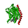 5ckrS S: Starting model for refinement C: citing same article ( |
|---|---|
| Similar structure data |

-Links
 PDBj
PDBj

- Assembly
| Deposited unit | 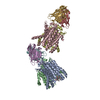
| ||||||||
|---|---|---|---|---|---|---|---|---|---|
| 1 | 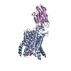
| ||||||||
| 2 | 
| ||||||||
| 3 | 
| ||||||||
| 4 | 
| ||||||||
| Unit cell |
|
-Components
| #1: Antibody | Mass: 15057.651 Da / Num. of mol.: 4 Source method: isolated from a genetically manipulated source Source: (gene. exp.) #2: Protein | Mass: 40954.684 Da / Num. of mol.: 4 Source method: isolated from a genetically manipulated source Source: (gene. exp.) Aquifex aeolicus (strain VF5) (bacteria)Strain: VF5 / Gene: mraY, aq_053 / Variant: VF5 / Production host: References: UniProt: O66465, phospho-N-acetylmuramoyl-pentapeptide-transferase #3: Chemical | ChemComp-NKM / ( |   Mass: 569.519 Da / Num. of mol.: 1 / Source method: obtained synthetically / Formula: C23H31N5O12 Mass: 569.519 Da / Num. of mol.: 1 / Source method: obtained synthetically / Formula: C23H31N5O12Has protein modification | Y | |
|---|
-Experimental details
-Experiment
| Experiment | Method: X-RAY DIFFRACTION / Number of used crystals: 1 |
|---|
- Sample preparation
| Crystal | Density Matthews: 3.36 Å3/Da / Density % sol: 63.42 % |
|---|---|
| Crystal grow | Temperature: 290 K / Method: vapor diffusion, sitting drop Details: 18% polyethylene glycol 4000, 0.4 M ammonium thiocyanate, 0.1 M sodium acetate pH 4.6 |
-Data collection
| Diffraction | Mean temperature: 100 K / Serial crystal experiment: N |
|---|---|
| Diffraction source | Source: SYNCHROTRON / Site: APS / Beamline: 24-ID-C / Wavelength: 0.9792 Å |
| Detector | Type: DECTRIS PILATUS 6M-F / Detector: PIXEL / Date: Mar 28, 2018 |
| Radiation | Protocol: SINGLE WAVELENGTH / Monochromatic (M) / Laue (L): M / Scattering type: x-ray |
| Radiation wavelength | Wavelength: 0.9792 Å / Relative weight: 1 |
| Reflection | Resolution: 3.62→87.64 Å / Num. obs: 32765 / % possible obs: 99.95 % / Redundancy: 15.2 % / CC1/2: 0.989 / Net I/σ(I): 10.25 |
| Reflection shell | Resolution: 3.62→3.749 Å / Num. unique obs: 3244 / CC1/2: 0.343 |
- Processing
| Software |
| |||||||||||||||||||||||||||||||||||||||||||||||||||||||||||||||||||||||||||||
|---|---|---|---|---|---|---|---|---|---|---|---|---|---|---|---|---|---|---|---|---|---|---|---|---|---|---|---|---|---|---|---|---|---|---|---|---|---|---|---|---|---|---|---|---|---|---|---|---|---|---|---|---|---|---|---|---|---|---|---|---|---|---|---|---|---|---|---|---|---|---|---|---|---|---|---|---|---|---|
| Refinement | Method to determine structure: MOLECULAR REPLACEMENT Starting model: 5CKR Resolution: 3.62→87.637 Å / SU ML: 0.64 / Cross valid method: FREE R-VALUE / σ(F): 1.33 / Phase error: 35.2
| |||||||||||||||||||||||||||||||||||||||||||||||||||||||||||||||||||||||||||||
| Solvent computation | Shrinkage radii: 0.9 Å / VDW probe radii: 1.11 Å | |||||||||||||||||||||||||||||||||||||||||||||||||||||||||||||||||||||||||||||
| Refinement step | Cycle: LAST / Resolution: 3.62→87.637 Å
| |||||||||||||||||||||||||||||||||||||||||||||||||||||||||||||||||||||||||||||
| Refine LS restraints |
| |||||||||||||||||||||||||||||||||||||||||||||||||||||||||||||||||||||||||||||
| LS refinement shell |
|
