 Movie
Movie Controller

[English] 日本語
 Yorodumi
- PDB-4xl4: Crystal structure of thiolase from Clostridium acetobutylicum in ... -
+ Open data

- Basic information
| Entry | Database: PDB / ID: 4xl4 | |||||||||
|---|---|---|---|---|---|---|---|---|---|---|
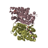
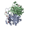
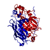
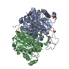
| Title | Crystal structure of thiolase from Clostridium acetobutylicum in complex with CoA | |||||||||
 Components Components | Acetyl-CoA acetyltransferase | |||||||||
 Keywords Keywords | TRANSFERASE | |||||||||
| Function / homology |  Function and homology information Function and homology informationacetyl-CoA C-acetyltransferase / acetyl-CoA C-acetyltransferase activity / cytoplasm Similarity search - Function | |||||||||
| Biological species |  Clostridium acetobutylicum (bacteria) Clostridium acetobutylicum (bacteria) | |||||||||
| Method |  X-RAY DIFFRACTION / SYNCHROTRON / MOLECULAR REPLACEMENT / Resolution: 1.9 Å X-RAY DIFFRACTION / SYNCHROTRON / MOLECULAR REPLACEMENT / Resolution: 1.9 Å | |||||||||
 Authors Authors | Kim, S. / Ha, S.C. / Ahn, J.W. / Kim, E.J. / Lim, J.H. / Kim, K.J. | |||||||||
 Citation Citation | Journal: Nat Commun / Year: 2015 Title: Redox-switch regulatory mechanism of thiolase from Clostridium acetobutylicum Authors: Kim, S. / Jang, Y.S. / Ha, S.C. / Ahn, J.W. / Kim, E.J. / Hong Lim, J. / Cho, C. / Shin Ryu, Y. / Kuk Lee, S. / Lee, S.Y. / Kim, K.J. | |||||||||
| History |
|
- Structure visualization
| Structure viewer | Molecule: MolmilJmol/JSmol |
|---|
- Downloads & links
-Download
| PDBx/mmCIF format | 4xl4.cif.gz | 175 KB | Display | PDBx/mmCIF format |
|---|---|---|---|---|
| PDB format | pdb4xl4.ent.gz | 136.7 KB | Display | PDB format |
| PDBx/mmJSON format | 4xl4.json.gz | Tree view | PDBx/mmJSON format | |
| Others |  Other downloads Other downloads |
-Validation report
| Arichive directory | https://data.pdbj.org/pub/pdb/validation_reports/xl/4xl4ftp://data.pdbj.org/pub/pdb/validation_reports/xl/4xl4 | HTTPS FTP |
|---|
-Related structure data
| Related structure data |  4wyrC  4wysC 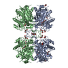 4xl2C  4xl3C 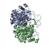 4n45S C: citing same article ( S: Starting model for refinement |
|---|---|
| Similar structure data |


-Links
 PDBj
PDBj


- Assembly
| Deposited unit | 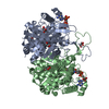
| ||||||||
|---|---|---|---|---|---|---|---|---|---|
| 1 |
| ||||||||
| Unit cell |
| ||||||||
| Details | biological unit is the same as asym. |
-Components
| #1: Protein | Mass: 42345.496 Da / Num. of mol.: 2 / Mutation: C378S Source method: isolated from a genetically manipulated source Source: (gene. exp.) Clostridium acetobutylicum (strain EA 2018) (bacteria)Strain: EA 2018 / Gene: CEA_G2880 / Plasmid: pET30a / Production host: References: UniProt: F0K5D8, UniProt: A0A0R4I970*PLUS, acetyl-CoA C-acetyltransferase #2: Chemical |   Mass: 767.534 Da / Num. of mol.: 2 / Source method: obtained synthetically / Formula: C21H36N7O16P3S Mass: 767.534 Da / Num. of mol.: 2 / Source method: obtained synthetically / Formula: C21H36N7O16P3S#3: Chemical | ChemComp-GOL /   Mass: 92.094 Da / Num. of mol.: 7 / Source method: obtained synthetically / Formula: C3H8O3 Mass: 92.094 Da / Num. of mol.: 7 / Source method: obtained synthetically / Formula: C3H8O3#4: Water | ChemComp-HOH / |  Mass: 18.015 Da / Num. of mol.: 636 / Source method: isolated from a natural source / Formula: H2O Mass: 18.015 Da / Num. of mol.: 636 / Source method: isolated from a natural source / Formula: H2O |
|---|
-Experimental details
-Experiment
| Experiment | Method: X-RAY DIFFRACTION / Number of used crystals: 1 |
|---|
- Sample preparation
| Crystal | Density Matthews: 2.45 Å3/Da / Density % sol: 49.81 % |
|---|---|
| Crystal grow | Temperature: 295 K / Method: vapor diffusion, hanging drop / pH: 4.2 / Details: PEG 3350, K-citrate, NaCl |
-Data collection
| Diffraction | Mean temperature: 100 K |
|---|---|
| Diffraction source | Source: SYNCHROTRON / Site: PAL/PLS  / Beamline: 7A (6B, 6C1) / Wavelength: 0.97934 Å / Beamline: 7A (6B, 6C1) / Wavelength: 0.97934 Å |
| Detector | Type: ADSC QUANTUM 270 / Detector: CCD / Date: Dec 23, 2014 |
| Radiation | Monochromator: DOUBLE CRYSTAL / Protocol: SINGLE WAVELENGTH / Monochromatic (M) / Laue (L): M / Scattering type: x-ray |
| Radiation wavelength | Wavelength: 0.97934 Å / Relative weight: 1 |
| Reflection | Resolution: 1.9→50 Å / Num. obs: 65291 / % possible obs: 99.9 % / Redundancy: 7 % / Rmerge(I) obs: 0.091 / Net I/σ(I): 33.1 |
| Reflection shell | Resolution: 1.9→1.93 Å / Redundancy: 6 % / Rmerge(I) obs: 0.282 / Mean I/σ(I) obs: 5.6 / Rejects: 0 / % possible all: 99.9 |
- Processing
| Software |
| |||||||||||||||||||||||||||||||||||||||||||||||||||||||||||||||||||||||||||
|---|---|---|---|---|---|---|---|---|---|---|---|---|---|---|---|---|---|---|---|---|---|---|---|---|---|---|---|---|---|---|---|---|---|---|---|---|---|---|---|---|---|---|---|---|---|---|---|---|---|---|---|---|---|---|---|---|---|---|---|---|---|---|---|---|---|---|---|---|---|---|---|---|---|---|---|---|
| Refinement | Method to determine structure: MOLECULAR REPLACEMENT Starting model: 4N45 Resolution: 1.9→50 Å / Cor.coef. Fo:Fc: 0.969 / Cor.coef. Fo:Fc free: 0.952 / WRfactor Rfree: 0.1776 / WRfactor Rwork: 0.1383 / FOM work R set: 0.9105 / SU B: 2.525 / SU ML: 0.075 / SU R Cruickshank DPI: 0.1177 / SU Rfree: 0.1155 / Cross valid method: THROUGHOUT / σ(F): 0 / ESU R: 0.119 / ESU R Free: 0.115 / Stereochemistry target values: MAXIMUM LIKELIHOOD Details: HYDROGENS HAVE BEEN ADDED IN THE RIDING POSITIONS U VALUES : REFINED INDIVIDUALLY
| |||||||||||||||||||||||||||||||||||||||||||||||||||||||||||||||||||||||||||
| Solvent computation | Ion probe radii: 0.8 Å / Shrinkage radii: 0.8 Å / VDW probe radii: 1.2 Å / Solvent model: MASK | |||||||||||||||||||||||||||||||||||||||||||||||||||||||||||||||||||||||||||
| Displacement parameters | Biso max: 102.3 Å2 / Biso mean: 24.167 Å2 / Biso min: 10.98 Å2
| |||||||||||||||||||||||||||||||||||||||||||||||||||||||||||||||||||||||||||
| Refinement step | Cycle: final / Resolution: 1.9→50 Å
| |||||||||||||||||||||||||||||||||||||||||||||||||||||||||||||||||||||||||||
| Refine LS restraints |
| |||||||||||||||||||||||||||||||||||||||||||||||||||||||||||||||||||||||||||
| LS refinement shell | Resolution: 1.897→1.946 Å / Total num. of bins used: 20
|
