 Movie
Movie Controller

[English] 日本語
 Yorodumi
- PDB-6oyu: Structure of an ancestral-reconstructed cytochrome P450 1B1 with ... -
+ Open data

- Basic information
| Entry | Database: PDB / ID: 6oyu | ||||||
|---|---|---|---|---|---|---|---|
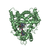
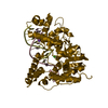
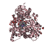
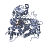
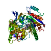
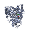
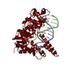
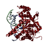
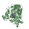
| Title | Structure of an ancestral-reconstructed cytochrome P450 1B1 with alpha-naphthoflavone | ||||||
 Components Components | Cytochrome P450 1B1 | ||||||
 Keywords Keywords | OXIDOREDUCTASE / CYP1B1 / ancestral reconstruction / mammalian | ||||||
| Function / homology | Cytochrome p450 / Cytochrome P450 / Orthogonal Bundle / Mainly Alpha / 2-PHENYL-4H-BENZO[H]CHROMEN-4-ONE / PROTOPORPHYRIN IX CONTAINING FE Function and homology information Function and homology information | ||||||
| Biological species | synthetic construct (others) | ||||||
| Method |  X-RAY DIFFRACTION / SYNCHROTRON / MOLECULAR REPLACEMENT / Resolution: 2.95 Å X-RAY DIFFRACTION / SYNCHROTRON / MOLECULAR REPLACEMENT / Resolution: 2.95 Å | ||||||
 Authors Authors | Bart, A.G. / Harris, K.L. / Scott, E.E. | ||||||
| Funding support |  United States, 1items United States, 1items
| ||||||
 Citation Citation | Journal: J.Biol.Chem. / Year: 2020 Title: Structure of an ancestral mammalian family 1B1 cytochrome P450 with increased thermostability. Authors: Bart, A.G. / Harris, K.L. / Gillam, E.M.J. / Scott, E.E. | ||||||
| History |
|
- Structure visualization
| Structure viewer | Molecule: MolmilJmol/JSmol |
|---|
- Downloads & links
-Download
| PDBx/mmCIF format | 6oyu.cif.gz | 359.7 KB | Display | PDBx/mmCIF format |
|---|---|---|---|---|
| PDB format | pdb6oyu.ent.gz | 296.8 KB | Display | PDB format |
| PDBx/mmJSON format | 6oyu.json.gz | Tree view | PDBx/mmJSON format | |
| Others |  Other downloads Other downloads |
-Validation report
| Arichive directory | https://data.pdbj.org/pub/pdb/validation_reports/oy/6oyuftp://data.pdbj.org/pub/pdb/validation_reports/oy/6oyu | HTTPS FTP |
|---|
-Related structure data
| Related structure data | 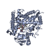 6oyvC  3pm0S S: Starting model for refinement C: citing same article ( |
|---|---|
| Similar structure data |

-Links
 PDBj
PDBj- Assembly
| Deposited unit | 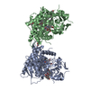
| ||||||||
|---|---|---|---|---|---|---|---|---|---|
| 1 | 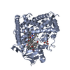
| ||||||||
| 2 | 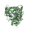
| ||||||||
| Unit cell |
|
-Components
| #1: Protein | Mass: 55349.980 Da / Num. of mol.: 2 Source method: isolated from a genetically manipulated source Source: (gene. exp.) synthetic construct (others) / Production host:  #2: Chemical | ChemComp-BHF /   Mass: 272.297 Da / Num. of mol.: 8 / Source method: obtained synthetically / Formula: C19H12O2 / Feature type: SUBJECT OF INVESTIGATION / Comment: inhibitor*YM Mass: 272.297 Da / Num. of mol.: 8 / Source method: obtained synthetically / Formula: C19H12O2 / Feature type: SUBJECT OF INVESTIGATION / Comment: inhibitor*YM#3: Chemical |   Mass: 616.487 Da / Num. of mol.: 2 / Source method: obtained synthetically / Formula: C34H32FeN4O4 Mass: 616.487 Da / Num. of mol.: 2 / Source method: obtained synthetically / Formula: C34H32FeN4O4#4: Chemical | ChemComp-GOL /   Mass: 92.094 Da / Num. of mol.: 6 / Source method: obtained synthetically / Formula: C3H8O3 Mass: 92.094 Da / Num. of mol.: 6 / Source method: obtained synthetically / Formula: C3H8O3#5: Water | ChemComp-HOH / |  Mass: 18.015 Da / Num. of mol.: 44 / Source method: isolated from a natural source / Formula: H2O Mass: 18.015 Da / Num. of mol.: 44 / Source method: isolated from a natural source / Formula: H2O |
|---|
-Experimental details
-Experiment
| Experiment | Method: X-RAY DIFFRACTION / Number of used crystals: 1 |
|---|
- Sample preparation
| Crystal | Density Matthews: 2.92 Å3/Da / Density % sol: 57.82 % |
|---|---|
| Crystal grow | Temperature: 293 K / Method: vapor diffusion, sitting drop / pH: 7 Details: 900 mM sodium citrate tribasic, 90 mM Tris base / HCl pH 7.0, 180 mM NaCl, 10% glycerol |
-Data collection
| Diffraction | Mean temperature: 100 K / Serial crystal experiment: N |
|---|---|
| Diffraction source | Source: SYNCHROTRON / Site: APS / Beamline: 21-ID-G / Wavelength: 0.9786 Å |
| Detector | Type: MARMOSAIC 300 mm CCD / Detector: CCD / Date: Oct 28, 2018 |
| Radiation | Protocol: SINGLE WAVELENGTH / Monochromatic (M) / Laue (L): M / Scattering type: x-ray |
| Radiation wavelength | Wavelength: 0.9786 Å / Relative weight: 1 |
| Reflection | Resolution: 2.95→50 Å / Num. obs: 27530 / % possible obs: 100 % / Redundancy: 7.5 % / CC1/2: 0.998 / Rpim(I) all: 0.064 / Net I/σ(I): 53 |
| Reflection shell | Resolution: 2.95→3 Å / Mean I/σ(I) obs: 5 / Num. unique obs: 1364 / CC1/2: 0.399 / Rpim(I) all: 0.788 |
- Processing
| Software |
| |||||||||||||||||||||||||||||||||||||||||||||||||||||||||||||||||||||||||||||||||||||||||||||||||||||||||
|---|---|---|---|---|---|---|---|---|---|---|---|---|---|---|---|---|---|---|---|---|---|---|---|---|---|---|---|---|---|---|---|---|---|---|---|---|---|---|---|---|---|---|---|---|---|---|---|---|---|---|---|---|---|---|---|---|---|---|---|---|---|---|---|---|---|---|---|---|---|---|---|---|---|---|---|---|---|---|---|---|---|---|---|---|---|---|---|---|---|---|---|---|---|---|---|---|---|---|---|---|---|---|---|---|---|---|
| Refinement | Method to determine structure: MOLECULAR REPLACEMENT Starting model: 3PM0 Resolution: 2.95→48.15 Å / SU ML: 0.33 / Cross valid method: FREE R-VALUE / σ(F): 1.35 / Phase error: 27.1
| |||||||||||||||||||||||||||||||||||||||||||||||||||||||||||||||||||||||||||||||||||||||||||||||||||||||||
| Solvent computation | Shrinkage radii: 0.9 Å / VDW probe radii: 1.11 Å | |||||||||||||||||||||||||||||||||||||||||||||||||||||||||||||||||||||||||||||||||||||||||||||||||||||||||
| Refinement step | Cycle: LAST / Resolution: 2.95→48.15 Å
| |||||||||||||||||||||||||||||||||||||||||||||||||||||||||||||||||||||||||||||||||||||||||||||||||||||||||
| Refine LS restraints |
| |||||||||||||||||||||||||||||||||||||||||||||||||||||||||||||||||||||||||||||||||||||||||||||||||||||||||
| LS refinement shell |
|
