 Movie
Movie Controller


+ Open data

- Basic information
| Entry | Database: PDB / ID: 6orb | |||||||||
|---|---|---|---|---|---|---|---|---|---|---|
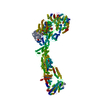
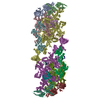
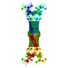
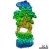
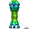
| Title | Full-length S. pombe Mdn1 in the presence of ATP and Rbin-1 | |||||||||
 Components Components | Midasin | |||||||||
 Keywords Keywords | MOTOR PROTEIN / Ribosome biogenesis / AAA protein / Mechanochemical enzyme / Ribosome assembly factor | |||||||||
| Function / homology |  Function and homology information Function and homology informationrixosome complex / ribosome biogenesis / ribosomal large subunit assembly / calcium ion binding / nucleolus / ATP hydrolysis activity / nucleoplasm / ATP binding / nucleus Similarity search - Function | |||||||||
| Biological species |  | |||||||||
| Method | ELECTRON MICROSCOPY / single particle reconstruction / cryo EM / Resolution: 7.7 Å | |||||||||
 Authors Authors | Chen, Z. / Suzuki, H. / Wang, A.C. / DiMaio, F. / Walz, T. / Kapoor, T.M. | |||||||||
| Funding support |  United States, 1items United States, 1items
| |||||||||
 Citation Citation | Journal: Cell / Year: 2018 Title: Structural Insights into Mdn1, an Essential AAA Protein Required for Ribosome Biogenesis. Authors: Zhen Chen / Hiroshi Suzuki / Yuki Kobayashi / Ashley C Wang / Frank DiMaio / Shigehiro A Kawashima / Thomas Walz / Tarun M Kapoor /  Abstract: Mdn1 is an essential AAA (ATPase associated with various activities) protein that removes assembly factors from distinct precursors of the ribosomal 60S subunit. However, Mdn1's large size (∼5,000 ...Mdn1 is an essential AAA (ATPase associated with various activities) protein that removes assembly factors from distinct precursors of the ribosomal 60S subunit. However, Mdn1's large size (∼5,000 amino acid [aa]) and its limited homology to other well-studied proteins have restricted our understanding of its remodeling function. Here, we present structures for S. pombe Mdn1 in the presence of AMPPNP at up to ∼4 Å or ATP plus Rbin-1, a chemical inhibitor, at ∼8 Å resolution. These data reveal that Mdn1's MIDAS domain is tethered to its ring-shaped AAA domain through an ∼20 nm long structured linker and a flexible ∼500 aa Asp/Glu-rich motif. We find that the MIDAS domain, which also binds other ribosome-assembly factors, docks onto the AAA ring in a nucleotide state-specific manner. Together, our findings reveal how conformational changes in the AAA ring can be directly transmitted to the MIDAS domain and thereby drive the targeted release of assembly factors from ribosomal 60S-subunit precursors. | |||||||||
| History |
|
- Structure visualization
| Movie |
Movie viewer |
|---|---|
| Structure viewer | Molecule: MolmilJmol/JSmol |
- Downloads & links
-Download
| PDBx/mmCIF format | 6orb.cif.gz | 495.4 KB | Display | PDBx/mmCIF format |
|---|---|---|---|---|
| PDB format | pdb6orb.ent.gz | 308.1 KB | Display | PDB format |
| PDBx/mmJSON format | 6orb.json.gz | Tree view | PDBx/mmJSON format | |
| Others |  Other downloads Other downloads |
-Validation report
| Arichive directory | https://data.pdbj.org/pub/pdb/validation_reports/or/6orbftp://data.pdbj.org/pub/pdb/validation_reports/or/6orb | HTTPS FTP |
|---|
-Related structure data
| Related structure data | 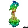 9036MC 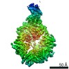 9032C 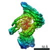 9033C 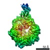 9034C 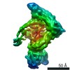 9035C 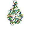 6or5C 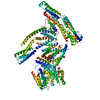 6or6C M: map data used to model this data C: citing same article ( |
|---|---|
| Similar structure data |


-Links
 PDBj
PDBj


- Assembly
| Deposited unit | 
|
|---|---|
| 1 |
|
-Components
| #1: Protein | Mass: 538381.125 Da / Num. of mol.: 1 Source method: isolated from a genetically manipulated source Source: (gene. exp.) Gene: mdn1, SPCC737.08 / Production host:  Trichoplusia ni (cabbage looper) / References: UniProt: O94248 Trichoplusia ni (cabbage looper) / References: UniProt: O94248 |
|---|
-Experimental details
-Experiment
| Experiment | Method: ELECTRON MICROSCOPY |
|---|---|
| EM experiment | Aggregation state: PARTICLE / 3D reconstruction method: single particle reconstruction |
- Sample preparation
| Component | Name: Full-length Mdn1 in the presence of ATP and Rbin-1 / Type: COMPLEX / Entity ID: all / Source: RECOMBINANT |
|---|---|
| Molecular weight | Value: 0.54 MDa / Experimental value: NO |
| Source (natural) | Organism: |
| Source (recombinant) | Organism: Trichoplusia ni (cabbage looper) |
| Buffer solution | pH: 7.5 |
| Specimen | Conc.: 0.15 mg/ml / Embedding applied: NO / Shadowing applied: NO / Staining applied: NO / Vitrification applied: YES |
| Specimen support | Grid material: COPPER / Grid mesh size: 400 divisions/in. / Grid type: Quantifoil R1.2/1.3 |
| Vitrification | Instrument: GATAN CRYOPLUNGE 3 / Cryogen name: ETHANE / Humidity: 80 % / Chamber temperature: 295 K |
- Electron microscopy imaging
| Experimental equipment |  Model: Talos Arctica / Image courtesy: FEI Company |
|---|---|
| Microscopy | Model: FEI TALOS ARCTICA |
| Electron gun | Electron source:  FIELD EMISSION GUN / Accelerating voltage: 200 kV / Illumination mode: FLOOD BEAM FIELD EMISSION GUN / Accelerating voltage: 200 kV / Illumination mode: FLOOD BEAM |
| Electron lens | Mode: BRIGHT FIELD / Nominal magnification: 28000 X / Nominal defocus max: 4000 nm / Nominal defocus min: 2000 nm / Cs: 2.7 mm / C2 aperture diameter: 70 µm |
| Specimen holder | Cryogen: NITROGEN / Specimen holder model: FEI TITAN KRIOS AUTOGRID HOLDER |
| Image recording | Average exposure time: 15 sec. / Electron dose: 66.7 e/Å2 / Detector mode: SUPER-RESOLUTION / Film or detector model: GATAN K2 SUMMIT (4k x 4k) / Num. of real images: 3887 |
| Image scans | Movie frames/image: 50 / Used frames/image: 1-50 |
- Processing
| EM software |
| ||||||||||||||||||||||||||||||||||||
|---|---|---|---|---|---|---|---|---|---|---|---|---|---|---|---|---|---|---|---|---|---|---|---|---|---|---|---|---|---|---|---|---|---|---|---|---|---|
| CTF correction | Type: PHASE FLIPPING AND AMPLITUDE CORRECTION | ||||||||||||||||||||||||||||||||||||
| Symmetry | Point symmetry: C1 (asymmetric) | ||||||||||||||||||||||||||||||||||||
| 3D reconstruction | Resolution: 7.7 Å / Resolution method: FSC 0.143 CUT-OFF / Num. of particles: 54557 / Symmetry type: POINT | ||||||||||||||||||||||||||||||||||||
| Atomic model building | Protocol: RIGID BODY FIT / Space: REAL |
