 Movie
Movie Controller

[English] 日本語
 Yorodumi
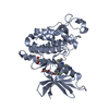
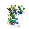
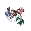
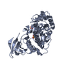
- PDB-6oni: Crystal structure of PPARgamma ligand binding domain in complex w... -
+ Open data

- Basic information
| Entry | Database: PDB / ID: 6oni | ||||||
|---|---|---|---|---|---|---|---|
| Title | Crystal structure of PPARgamma ligand binding domain in complex with N-CoR peptide and inverse agonist T0070907 | ||||||
 Components Components |
| ||||||
 Keywords Keywords | transcription/agonist / Nuclear receptors / TZDs / Drug design / Therapeutic targets / TRANSCRIPTION / transcription-agonist complex | ||||||
| Function / homology |  Function and homology information Function and homology informationLoss of MECP2 binding ability to the NCoR/SMRT complex / negative regulation of androgen receptor signaling pathway / prostaglandin receptor activity / negative regulation of receptor signaling pathway via STAT / MECP2 regulates transcription factors / beige fat cell differentiation / negative regulation of vascular endothelial cell proliferation / negative regulation of extracellular matrix assembly / negative regulation of connective tissue replacement involved in inflammatory response wound healing / nuclear thyroid hormone receptor binding ...Loss of MECP2 binding ability to the NCoR/SMRT complex / negative regulation of androgen receptor signaling pathway / prostaglandin receptor activity / negative regulation of receptor signaling pathway via STAT / MECP2 regulates transcription factors / beige fat cell differentiation / negative regulation of vascular endothelial cell proliferation / negative regulation of extracellular matrix assembly / negative regulation of connective tissue replacement involved in inflammatory response wound healing / nuclear thyroid hormone receptor binding / positive regulation of cholesterol transport / negative regulation of cellular response to transforming growth factor beta stimulus / negative regulation of glycolytic process / negative regulation of JNK cascade / arachidonate binding / positive regulation of adiponectin secretion / NR1H2 & NR1H3 regulate gene expression to control bile acid homeostasis / DNA binding domain binding / negative regulation of cardiac muscle hypertrophy in response to stress / positive regulation of vascular associated smooth muscle cell apoptotic process / negative regulation of fatty acid metabolic process / positive regulation of lipid metabolic process / positive regulation of fatty acid metabolic process / STAT family protein binding / Notch-HLH transcription pathway / WW domain binding / negative regulation of type II interferon-mediated signaling pathway / LBD domain binding / negative regulation of cholesterol storage / locomotor rhythm / response to lipid / positive regulation of lipoprotein transport / negative regulation of SMAD protein signal transduction / histone deacetylase complex / lipid homeostasis / E-box binding / R-SMAD binding / negative regulation of blood vessel endothelial cell migration / white fat cell differentiation / negative regulation of vascular associated smooth muscle cell proliferation / alpha-actinin binding / negative regulation of macrophage derived foam cell differentiation / negative regulation of lipid storage / positive regulation of cholesterol efflux / monocyte differentiation / negative regulation of BMP signaling pathway / Regulation of MECP2 expression and activity / cell fate commitment / cellular response to low-density lipoprotein particle stimulus / long-chain fatty acid transport / BMP signaling pathway / negative regulation of mitochondrial fission / Nuclear signaling by ERBB4 / negative regulation of osteoblast differentiation / positive regulation of fat cell differentiation / nuclear retinoid X receptor binding / fat cell differentiation / Transcriptional regulation of brown and beige adipocyte differentiation by EBF2 / retinoic acid receptor signaling pathway / NR1H3 & NR1H2 regulate gene expression linked to cholesterol transport and efflux / intracellular receptor signaling pathway / Regulation of lipid metabolism by PPARalpha / negative regulation of MAPK cascade / spindle assembly / transcription repressor complex / cell maturation / peptide binding / peroxisome proliferator activated receptor signaling pathway / epithelial cell differentiation / hormone-mediated signaling pathway / regulation of cellular response to insulin stimulus / positive regulation of adipose tissue development / response to nutrient / RORA,B,C and NR1D1 (REV-ERBA) regulate gene expression / Expression of BMAL (ARNTL), CLOCK, and NPAS2 / negative regulation of miRNA transcription / negative regulation of angiogenesis / brown fat cell differentiation / placenta development / nuclear receptor binding / Regulation of PTEN gene transcription / transcription coregulator binding / SUMOylation of intracellular receptors / positive regulation of apoptotic signaling pathway / negative regulation of smooth muscle cell proliferation / HDACs deacetylate histones / Heme signaling / negative regulation of transforming growth factor beta receptor signaling pathway / PPARA activates gene expression / Transcriptional activation of mitochondrial biogenesis / Cytoprotection by HMOX1 / Downregulation of SMAD2/3:SMAD4 transcriptional activity / fatty acid metabolic process / Nuclear Receptor transcription pathway / Transcriptional regulation of white adipocyte differentiation / regulation of circadian rhythm / positive regulation of miRNA transcription / mRNA transcription by RNA polymerase II / DNA-binding transcription repressor activity, RNA polymerase II-specific / NOTCH1 Intracellular Domain Regulates Transcription Similarity search - Function | ||||||
| Biological species |  Homo sapiens (human) Homo sapiens (human) | ||||||
| Method |  X-RAY DIFFRACTION / SYNCHROTRON / MOLECULAR REPLACEMENT / molecular replacement / Resolution: 1.8 Å X-RAY DIFFRACTION / SYNCHROTRON / MOLECULAR REPLACEMENT / molecular replacement / Resolution: 1.8 Å | ||||||
 Authors Authors | Shang, J. / Kojetin, D.J. | ||||||
| Funding support |  United States, 1items United States, 1items
| ||||||
 Citation Citation | Journal: Nat Commun / Year: 2020 Title: A molecular switch regulating transcriptional repression and activation of PPAR gamma. Authors: Shang, J. / Mosure, S.A. / Zheng, J. / Brust, R. / Bass, J. / Nichols, A. / Solt, L.A. / Griffin, P.R. / Kojetin, D.J. | ||||||
| History |
|
- Structure visualization
| Structure viewer | Molecule: MolmilJmol/JSmol |
|---|
- Downloads & links
-Download
| PDBx/mmCIF format | 6oni.cif.gz | 81 KB | Display | PDBx/mmCIF format |
|---|---|---|---|---|
| PDB format | pdb6oni.ent.gz | 57.4 KB | Display | PDB format |
| PDBx/mmJSON format | 6oni.json.gz | Tree view | PDBx/mmJSON format | |
| Others |  Other downloads Other downloads |
-Validation report
| Arichive directory | https://data.pdbj.org/pub/pdb/validation_reports/on/6oniftp://data.pdbj.org/pub/pdb/validation_reports/on/6oni | HTTPS FTP |
|---|
-Related structure data
| Related structure data |  6onjC  6pdzC  6c1iS S: Starting model for refinement C: citing same article ( |
|---|---|
| Similar structure data |


-Links
 PDBj
PDBj


- Assembly
| Deposited unit | 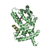
| ||||||||
|---|---|---|---|---|---|---|---|---|---|
| 1 |
| ||||||||
| Unit cell |
|
-Components
| #1: Protein | Mass: 31449.520 Da / Num. of mol.: 1 Source method: isolated from a genetically manipulated source Source: (gene. exp.) Homo sapiens (human) / Gene: PPARG, NR1C3 / Plasmid: pET46 / Production host:  |
|---|---|
| #2: Protein/peptide | Mass: 2508.824 Da / Num. of mol.: 1 Source method: isolated from a genetically manipulated source Source: (gene. exp.) Homo sapiens (human) / Production host: |
| #3: Chemical | ChemComp-EEY / 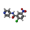  Mass: 277.663 Da / Num. of mol.: 1 / Source method: obtained synthetically / Formula: C12H8ClN3O3 / Feature type: SUBJECT OF INVESTIGATION Mass: 277.663 Da / Num. of mol.: 1 / Source method: obtained synthetically / Formula: C12H8ClN3O3 / Feature type: SUBJECT OF INVESTIGATION |
| #4: Water | ChemComp-HOH /  Mass: 18.015 Da / Num. of mol.: 391 / Source method: isolated from a natural source / Formula: H2O Mass: 18.015 Da / Num. of mol.: 391 / Source method: isolated from a natural source / Formula: H2O |
| Has ligand of interest | Y |
| Has protein modification | Y |
-Experimental details
-Experiment
| Experiment | Method: X-RAY DIFFRACTION / Number of used crystals: 1 |
|---|
- Sample preparation
| Crystal | Density Matthews: 2.32 Å3/Da / Density % sol: 46.91 % |
|---|---|
| Crystal grow | Temperature: 293 K / Method: vapor diffusion, sitting drop / pH: 6.5 Details: 0.2M Ammonium sulfate 0.1M MES, pH 6.5 30% w/v, PEG 8000 |
-Data collection
| Diffraction | Mean temperature: 100 K / Serial crystal experiment: N |
|---|---|
| Diffraction source | Source: SYNCHROTRON / Site: SSRL / Beamline: BL12-2 / Wavelength: 0.97946 Å |
| Detector | Type: DECTRIS PILATUS 6M / Detector: PIXEL / Date: Jan 17, 2019 |
| Radiation | Monochromator: Liquid nitrogen-cooled double crystal Si(111) Protocol: SINGLE WAVELENGTH / Monochromatic (M) / Laue (L): M / Scattering type: x-ray |
| Radiation wavelength | Wavelength: 0.97946 Å / Relative weight: 1 |
| Reflection | Resolution: 1.8→34.233 Å / Num. obs: 30550 / % possible obs: 99.49 % / Redundancy: 2 % / Biso Wilson estimate: 25.96 Å2 / CC1/2: 1 / Rmerge(I) obs: 0.01592 / Rpim(I) all: 0.01592 / Rrim(I) all: 0.02251 / Net I/σ(I): 21.08 |
| Reflection shell | Resolution: 1.8→1.864 Å / Redundancy: 2 % / Rmerge(I) obs: 0.138 / Mean I/σ(I) obs: 3.29 / Num. unique obs: 2995 / CC1/2: 0.956 / Rpim(I) all: 0.138 / Rrim(I) all: 0.1952 / % possible all: 99.93 |
-Phasing
| Phasing | Method: molecular replacement |
|---|
- Processing
| Software |
| ||||||||||||||||||||||||||||||||||||||||||||||||||||||||||||||||||||||||||||||||||||||||||
|---|---|---|---|---|---|---|---|---|---|---|---|---|---|---|---|---|---|---|---|---|---|---|---|---|---|---|---|---|---|---|---|---|---|---|---|---|---|---|---|---|---|---|---|---|---|---|---|---|---|---|---|---|---|---|---|---|---|---|---|---|---|---|---|---|---|---|---|---|---|---|---|---|---|---|---|---|---|---|---|---|---|---|---|---|---|---|---|---|---|---|---|
| Refinement | Method to determine structure: MOLECULAR REPLACEMENT Starting model: 6C1I Resolution: 1.8→34.233 Å / SU ML: 0.2 / Cross valid method: THROUGHOUT / σ(F): 1.34 / Phase error: 23.24 / Stereochemistry target values: ML
| ||||||||||||||||||||||||||||||||||||||||||||||||||||||||||||||||||||||||||||||||||||||||||
| Solvent computation | Shrinkage radii: 0.9 Å / VDW probe radii: 1.11 Å / Solvent model: FLAT BULK SOLVENT MODEL | ||||||||||||||||||||||||||||||||||||||||||||||||||||||||||||||||||||||||||||||||||||||||||
| Displacement parameters | Biso max: 74.11 Å2 / Biso mean: 29.5468 Å2 / Biso min: 9.71 Å2 | ||||||||||||||||||||||||||||||||||||||||||||||||||||||||||||||||||||||||||||||||||||||||||
| Refinement step | Cycle: final / Resolution: 1.8→34.233 Å
| ||||||||||||||||||||||||||||||||||||||||||||||||||||||||||||||||||||||||||||||||||||||||||
| Refine LS restraints |
| ||||||||||||||||||||||||||||||||||||||||||||||||||||||||||||||||||||||||||||||||||||||||||
| LS refinement shell | Refine-ID: X-RAY DIFFRACTION / Rfactor Rfree error: 0
|
