 Movie
Movie Controller

[English] 日本語
 Yorodumi
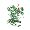
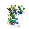
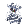
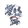
- PDB-6f87: Crystal structure of P. abyssi Sua5 complexed with L-threonine and PPi -
+ Open data

- Basic information
| Entry | Database: PDB / ID: 6f87 | ||||||
|---|---|---|---|---|---|---|---|
| Title | Crystal structure of P. abyssi Sua5 complexed with L-threonine and PPi | ||||||
 Components Components | Threonylcarbamoyl-AMP synthase | ||||||
 Keywords Keywords | TRANSFERASE / Nucleotidyltransferase / tRNA modification / threonylcarbamoylation | ||||||
| Function / homology |  Function and homology information Function and homology informationL-threonylcarbamoyladenylate synthase / L-threonylcarbamoyladenylate synthase activity / tRNA threonylcarbamoyladenosine modification / regulation of translational fidelity / double-stranded RNA binding / ATP binding / cytoplasm Similarity search - Function | ||||||
| Biological species |   Pyrococcus abyssi (archaea) Pyrococcus abyssi (archaea) | ||||||
| Method |  X-RAY DIFFRACTION / SYNCHROTRON / MOLECULAR REPLACEMENT / Resolution: 2.62 Å X-RAY DIFFRACTION / SYNCHROTRON / MOLECULAR REPLACEMENT / Resolution: 2.62 Å | ||||||
 Authors Authors | Pichard-Kostuch, A. / Zhang, W. / Liger, D. / Daugeron, M.C. / Letoquart, J. / Li de la Sierra-Gallay, I. / Forterre, P. / Collinet, B. / van Tilbeurgh, H. / Basta, T. | ||||||
| Funding support |  France, 1items France, 1items
| ||||||
 Citation Citation | Journal: RNA / Year: 2018 Title: Structure-function analysis of Sua5 protein reveals novel functional motifs required for the biosynthesis of the universal t6A tRNA modification. Authors: Pichard-Kostuch, A. / Zhang, W. / Liger, D. / Daugeron, M.C. / Letoquart, J. / Li de la Sierra-Gallay, I. / Forterre, P. / Collinet, B. / van Tilbeurgh, H. / Basta, T. | ||||||
| History |
|
- Structure visualization
| Structure viewer | Molecule: MolmilJmol/JSmol |
|---|
- Downloads & links
-Download
| PDBx/mmCIF format | 6f87.cif.gz | 269.8 KB | Display | PDBx/mmCIF format |
|---|---|---|---|---|
| PDB format | pdb6f87.ent.gz | 220.2 KB | Display | PDB format |
| PDBx/mmJSON format | 6f87.json.gz | Tree view | PDBx/mmJSON format | |
| Others |  Other downloads Other downloads |
-Validation report
| Arichive directory | https://data.pdbj.org/pub/pdb/validation_reports/f8/6f87ftp://data.pdbj.org/pub/pdb/validation_reports/f8/6f87 | HTTPS FTP |
|---|
-Related structure data
| Related structure data |  6f89C  6f8yC  2eqaS S: Starting model for refinement C: citing same article ( |
|---|---|
| Similar structure data |


-Links
 PDBj
PDBj


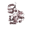
- Assembly
| Deposited unit | 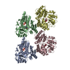
| |||||||||
|---|---|---|---|---|---|---|---|---|---|---|
| 1 | 
| |||||||||
| 2 | 
| |||||||||
| 3 | 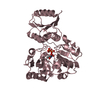
| |||||||||
| 4 | 
| |||||||||
| Unit cell |
| |||||||||
| Components on special symmetry positions |
|
-Components
| #1: Protein | Mass: 38197.156 Da / Num. of mol.: 4 Source method: isolated from a genetically manipulated source Source: (gene. exp.) Pyrococcus abyssi (strain GE5 / Orsay) (archaea)Strain: GE5 / Orsay / Gene: sua5, PYRAB15960, PAB1302 / Production host:  References: UniProt: Q9UYB2, L-threonylcarbamoyladenylate synthase #2: Chemical | ChemComp-POP /   Mass: 175.959 Da / Num. of mol.: 4 / Source method: isolated from a natural source / Formula: H2O7P2 Mass: 175.959 Da / Num. of mol.: 4 / Source method: isolated from a natural source / Formula: H2O7P2#3: Chemical | ChemComp-THR / 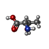  Type: L-peptide linking / Mass: 119.119 Da / Num. of mol.: 4 / Source method: obtained synthetically / Formula: C4H9NO3 Type: L-peptide linking / Mass: 119.119 Da / Num. of mol.: 4 / Source method: obtained synthetically / Formula: C4H9NO3#4: Water | ChemComp-HOH / |  Mass: 18.015 Da / Num. of mol.: 102 / Source method: isolated from a natural source / Formula: H2O Mass: 18.015 Da / Num. of mol.: 102 / Source method: isolated from a natural source / Formula: H2O |
|---|
-Experimental details
-Experiment
| Experiment | Method: X-RAY DIFFRACTION / Number of used crystals: 1 |
|---|
- Sample preparation
| Crystal | Density Matthews: 3.57 Å3/Da / Density % sol: 65.53 % |
|---|---|
| Crystal grow | Temperature: 277 K / Method: vapor diffusion, sitting drop / Details: ammonium sulfate / PH range: 8.5 - 8.8 |
-Data collection
| Diffraction | Mean temperature: 100 K |
|---|---|
| Diffraction source | Source: SYNCHROTRON / Site: SOLEIL / Beamline: PROXIMA 1 / Wavelength: 0.98011 Å |
| Detector | Type: DECTRIS PILATUS 6M / Detector: PIXEL / Date: Dec 4, 2011 |
| Radiation | Protocol: SINGLE WAVELENGTH / Monochromatic (M) / Laue (L): M / Scattering type: x-ray |
| Radiation wavelength | Wavelength: 0.98011 Å / Relative weight: 1 |
| Reflection | Resolution: 2.62→49.03 Å / Num. obs: 64452 / % possible obs: 99.4 % / Redundancy: 3.8 % / Rrim(I) all: 0.071 / Net I/σ(I): 14.14 |
| Reflection shell | Resolution: 2.62→2.78 Å / Redundancy: 3.8 % / Mean I/σ(I) obs: 2.11 / Num. unique obs: 10212 / Rrim(I) all: 0.69 / % possible all: 97.7 |
- Processing
| Software |
| ||||||||||||||||||||||||||||||||||||||||||||||||||||||||||||
|---|---|---|---|---|---|---|---|---|---|---|---|---|---|---|---|---|---|---|---|---|---|---|---|---|---|---|---|---|---|---|---|---|---|---|---|---|---|---|---|---|---|---|---|---|---|---|---|---|---|---|---|---|---|---|---|---|---|---|---|---|---|
| Refinement | Method to determine structure: MOLECULAR REPLACEMENT Starting model: 2EQA Resolution: 2.62→49.03 Å / Cor.coef. Fo:Fc: 0.968 / Cor.coef. Fo:Fc free: 0.942 / SU B: 10.58 / SU ML: 0.214 / Cross valid method: THROUGHOUT / σ(F): 0 / ESU R: 0.358 / ESU R Free: 0.258 / Stereochemistry target values: MAXIMUM LIKELIHOOD Details: HYDROGENS HAVE BEEN ADDED IN THE RIDING POSITIONS U VALUES : REFINED INDIVIDUALLY
| ||||||||||||||||||||||||||||||||||||||||||||||||||||||||||||
| Solvent computation | Ion probe radii: 0.8 Å / Shrinkage radii: 0.8 Å / VDW probe radii: 1.2 Å / Solvent model: MASK | ||||||||||||||||||||||||||||||||||||||||||||||||||||||||||||
| Displacement parameters | Biso max: 236.33 Å2 / Biso mean: 66.881 Å2 / Biso min: 35.85 Å2
| ||||||||||||||||||||||||||||||||||||||||||||||||||||||||||||
| Refinement step | Cycle: final / Resolution: 2.62→49.03 Å
| ||||||||||||||||||||||||||||||||||||||||||||||||||||||||||||
| Refine LS restraints |
| ||||||||||||||||||||||||||||||||||||||||||||||||||||||||||||
| LS refinement shell | Resolution: 2.617→2.685 Å / Rfactor Rfree error: 0 / Total num. of bins used: 20
|
