 Movie
Movie Controller

[English] 日本語
 Yorodumi
- PDB-6ofh: Structure of Salmonella type III secretion system needle filament -
+ Open data

- Basic information
| Entry | Database: PDB / ID: 6ofh | |||||||||||||||||||||||||||||||||
|---|---|---|---|---|---|---|---|---|---|---|---|---|---|---|---|---|---|---|---|---|---|---|---|---|---|---|---|---|---|---|---|---|---|---|
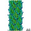
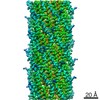
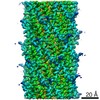
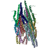
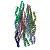
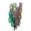
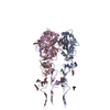
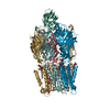
| Title | Structure of Salmonella type III secretion system needle filament | |||||||||||||||||||||||||||||||||
 Components Components | Protein PrgI | |||||||||||||||||||||||||||||||||
 Keywords Keywords | PROTEIN TRANSPORT / type III secretion system / salmonella / helical reconstruction | |||||||||||||||||||||||||||||||||
| Function / homology |  Function and homology information Function and homology informationtype III protein secretion system complex / protein secretion by the type III secretion system / cell surface / extracellular region / identical protein binding Similarity search - Function | |||||||||||||||||||||||||||||||||
| Biological species |  Salmonella typhimurium (bacteria) Salmonella typhimurium (bacteria) | |||||||||||||||||||||||||||||||||
| Method | ELECTRON MICROSCOPY / helical reconstruction / cryo EM / Resolution: 3.7 Å | |||||||||||||||||||||||||||||||||
 Authors Authors | Guo, E.Z. / Galan, J.E. | |||||||||||||||||||||||||||||||||
| Funding support |  United States, 1items United States, 1items
| |||||||||||||||||||||||||||||||||
 Citation Citation | Journal: PLoS Biol / Year: 2019 Title: A polymorphic helix of a Salmonella needle protein relays signals defining distinct steps in type III secretion. Authors: Emily Z Guo / Daniel C Desrosiers / Jan Zalesak / James Tolchard / Mélanie Berbon / Birgit Habenstein / Thomas Marlovits / Antoine Loquet / Jorge E Galán /    Abstract: Type III protein-secretion machines are essential for the interactions of many pathogenic or symbiotic bacterial species with their respective eukaryotic hosts. The core component of these machines ...Type III protein-secretion machines are essential for the interactions of many pathogenic or symbiotic bacterial species with their respective eukaryotic hosts. The core component of these machines is the injectisome, a multiprotein complex that mediates the selection of substrates, their passage through the bacterial envelope, and ultimately their delivery into eukaryotic target cells. The injectisome is composed of a large cytoplasmic complex or sorting platform, a multiring base embedded in the bacterial envelope, and a needle-like filament that protrudes several nanometers from the bacterial surface and is capped at its distal end by the tip complex. A characteristic feature of these machines is that their activity is stimulated by contact with target host cells. The sensing of target cells, thought to be mediated by the distal tip of the needle filament, generates an activating signal that must be transduced to the secretion machine by the needle filament. Here, through a multidisciplinary approach, including solid-state NMR (SSNMR) and cryo electron microscopy (cryo-EM) analyses, we have identified critical residues of the needle filament protein of a Salmonella Typhimurium type III secretion system that are involved in the regulation of the activity of the secretion machine. We found that mutations in the needle filament protein result in various specific phenotypes associated with different steps in the type III secretion process. More specifically, these studies reveal an important role for a polymorphic helix of the needle filament protein and the residues that line the lumen of its central channel in the control of type III secretion. | |||||||||||||||||||||||||||||||||
| History |
|
- Structure visualization
| Movie |
Movie viewer |
|---|---|
| Structure viewer | Molecule: MolmilJmol/JSmol |
- Downloads & links
-Download
| PDBx/mmCIF format | 6ofh.cif.gz | 252.7 KB | Display | PDBx/mmCIF format |
|---|---|---|---|---|
| PDB format | pdb6ofh.ent.gz | 210.6 KB | Display | PDB format |
| PDBx/mmJSON format | 6ofh.json.gz | Tree view | PDBx/mmJSON format | |
| Others |  Other downloads Other downloads |
-Validation report
| Summary document | 6ofh_validation.pdf.gz | 895.6 KB | Display | wwPDB validaton report |
|---|---|---|---|---|
| Full document | 6ofh_full_validation.pdf.gz | 903.5 KB | Display | |
| Data in XML | 6ofh_validation.xml.gz | 32.4 KB | Display | |
| Data in CIF | 6ofh_validation.cif.gz | 51.9 KB | Display | |
| Arichive directory | https://data.pdbj.org/pub/pdb/validation_reports/of/6ofhftp://data.pdbj.org/pub/pdb/validation_reports/of/6ofh | HTTPS FTP |
-Related structure data
| Related structure data | 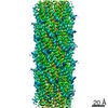 20046MC 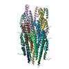 6ofeC 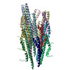 6offC 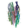 6ofgC M: map data used to model this data C: citing same article ( |
|---|---|
| Similar structure data |

-Links
 PDBj
PDBj- Assembly
| Deposited unit | 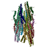
|
|---|---|
| 1 |
|
| Symmetry | Helical symmetry: (Circular symmetry: 1 / Dyad axis: no / N subunits divisor: 1 / Num. of operations: 19 / Rise per n subunits: 4.24 Å / Rotation per n subunits: 63.35 °) |
-Components
| #1: Protein | Mass: 8864.868 Da / Num. of mol.: 19 / Source method: isolated from a natural source Source: (natural) Salmonella typhimurium (strain SL1344) (bacteria)Strain: SL1344 / References: UniProt: A0A0H3NF82, UniProt: P41784*PLUS Has protein modification | N | |
|---|
-Experimental details
-Experiment
| Experiment | Method: ELECTRON MICROSCOPY |
|---|---|
| EM experiment | Aggregation state: FILAMENT / 3D reconstruction method: helical reconstruction |
- Sample preparation
| Component | Name: filament structure of PrgI needle attached to the base Type: COMPLEX / Entity ID: all / Source: NATURAL |
|---|---|
| Source (natural) | Organism: Salmonella typhimurium (strain SL1344) (bacteria) |
| Buffer solution | pH: 7.5 |
| Specimen | Embedding applied: NO / Shadowing applied: NO / Staining applied: NO / Vitrification applied: YES |
| Specimen support | Details: unspecified |
| Vitrification | Cryogen name: ETHANE |
- Electron microscopy imaging
| Experimental equipment |  Model: Titan Krios / Image courtesy: FEI Company |
|---|---|
| Microscopy | Model: FEI TITAN KRIOS |
| Electron gun | Electron source:  FIELD EMISSION GUN / Accelerating voltage: 300 kV / Illumination mode: FLOOD BEAM FIELD EMISSION GUN / Accelerating voltage: 300 kV / Illumination mode: FLOOD BEAM |
| Electron lens | Mode: BRIGHT FIELD |
| Image recording | Electron dose: 47 e/Å2 / Film or detector model: GATAN K2 SUMMIT (4k x 4k) |
- Processing
| Software | Name: PHENIX / Version: 1.12_2829: / Classification: refinement | ||||||||||||||||||||||||
|---|---|---|---|---|---|---|---|---|---|---|---|---|---|---|---|---|---|---|---|---|---|---|---|---|---|
| EM software | Name: PHENIX / Category: model refinement | ||||||||||||||||||||||||
| CTF correction | Type: PHASE FLIPPING AND AMPLITUDE CORRECTION | ||||||||||||||||||||||||
| Helical symmerty | Angular rotation/subunit: 63.35 ° / Axial rise/subunit: 4.24 Å / Axial symmetry: C1 | ||||||||||||||||||||||||
| 3D reconstruction | Resolution: 3.7 Å / Resolution method: FSC 0.143 CUT-OFF / Num. of particles: 53866 / Symmetry type: HELICAL | ||||||||||||||||||||||||
| Refine LS restraints |
|
