 Movie
Movie Controller

[English] 日本語
 Yorodumi
- PDB-6oeb: Crystal structure of HMCES SRAP domain in complex with 3' overhang DNA -
+ Open data

- Basic information
| Entry | Database: PDB / ID: 6oeb | |||||||||
|---|---|---|---|---|---|---|---|---|---|---|
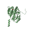
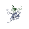
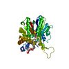
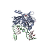
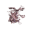
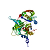
| Title | Crystal structure of HMCES SRAP domain in complex with 3' overhang DNA | |||||||||
 Components Components |
| |||||||||
 Keywords Keywords | DNA BINDING PROTEIN/DNA / SRAP Domain / HMCES / Structural Genomics / Structural Genomics Consortium / SGC / DNA binding protein / DNA-protein complex / DNA-damage protein / 3' overhang / DNA BINDING PROTEIN-DNA complex | |||||||||
| Function / homology |  Function and homology information Function and homology informationprotein-DNA covalent cross-linking activity / Lyases / DNA-(abasic site) binding / positive regulation of isotype switching / double-strand break repair via alternative nonhomologous end joining / protein-DNA covalent cross-linking repair / Hydrolases; Acting on peptide bonds (peptidases) / interstrand cross-link repair / somatic hypermutation of immunoglobulin genes / replication fork ...protein-DNA covalent cross-linking activity / Lyases / DNA-(abasic site) binding / positive regulation of isotype switching / double-strand break repair via alternative nonhomologous end joining / protein-DNA covalent cross-linking repair / Hydrolases; Acting on peptide bonds (peptidases) / interstrand cross-link repair / somatic hypermutation of immunoglobulin genes / replication fork / peptidase activity / single-stranded DNA binding / DNA damage response / proteolysis Similarity search - Function | |||||||||
| Biological species |  Homo sapiens (human) Homo sapiens (human)synthetic construct (others) | |||||||||
| Method |  X-RAY DIFFRACTION / SYNCHROTRON / MOLECULAR REPLACEMENT / Resolution: 2.1 Å X-RAY DIFFRACTION / SYNCHROTRON / MOLECULAR REPLACEMENT / Resolution: 2.1 Å | |||||||||
 Authors Authors | Halabelian, L. / Ravichandran, M. / Li, Y. / Zeng, H. / Bountra, C. / Edwards, A.M. / Arrowsmith, C.H. / Structural Genomics Consortium (SGC) | |||||||||
 Citation Citation | Journal: Nat.Struct.Mol.Biol. / Year: 2019 Title: Structural basis of HMCES interactions with abasic DNA and multivalent substrate recognition. Authors: Halabelian, L. / Ravichandran, M. / Li, Y. / Zeng, H. / Rao, A. / Aravind, L. / Arrowsmith, C.H. | |||||||||
| History |
|
- Structure visualization
| Structure viewer | Molecule: MolmilJmol/JSmol |
|---|
- Downloads & links
-Download
| PDBx/mmCIF format | 6oeb.cif.gz | 81.8 KB | Display | PDBx/mmCIF format |
|---|---|---|---|---|
| PDB format | pdb6oeb.ent.gz | 55.3 KB | Display | PDB format |
| PDBx/mmJSON format | 6oeb.json.gz | Tree view | PDBx/mmJSON format | |
| Others |  Other downloads Other downloads |
-Validation report
| Summary document | 6oeb_validation.pdf.gz | 450.3 KB | Display | wwPDB validaton report |
|---|---|---|---|---|
| Full document | 6oeb_full_validation.pdf.gz | 451.1 KB | Display | |
| Data in XML | 6oeb_validation.xml.gz | 12.7 KB | Display | |
| Data in CIF | 6oeb_validation.cif.gz | 17.4 KB | Display | |
| Arichive directory | https://data.pdbj.org/pub/pdb/validation_reports/oe/6oebftp://data.pdbj.org/pub/pdb/validation_reports/oe/6oeb | HTTPS FTP |
-Related structure data
| Related structure data |  5ko9SC  6oe7C  6oeaC S: Starting model for refinement C: citing same article ( |
|---|---|
| Similar structure data |


-Links
 PDBj
PDBj- Assembly
| Deposited unit | 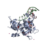
| ||||||||
|---|---|---|---|---|---|---|---|---|---|
| 1 | 
| ||||||||
| Unit cell |
|
-Components
-Protein , 1 types, 1 molecules A
| #1: Protein | Mass: 31671.617 Da / Num. of mol.: 1 / Fragment: SRAP domain (UNP residues 2-270) Source method: isolated from a genetically manipulated source Source: (gene. exp.) Homo sapiens (human) / Gene: HMCES, C3orf37, DC12, SRAPD1 / Production host:  |
|---|
-DNA chain , 2 types, 2 molecules BC
| #2: DNA chain | Mass: 2715.799 Da / Num. of mol.: 1 / Source method: obtained synthetically / Source: (synth.) synthetic construct (others) |
|---|---|
| #3: DNA chain | Mass: 1840.227 Da / Num. of mol.: 1 / Source method: obtained synthetically / Source: (synth.) synthetic construct (others) |
-Non-polymers , 3 types, 94 molecules 


| #4: Chemical | ChemComp-EDO /  Mass: 62.068 Da / Num. of mol.: 6 / Source method: obtained synthetically / Formula: C2H6O2 Mass: 62.068 Da / Num. of mol.: 6 / Source method: obtained synthetically / Formula: C2H6O2#5: Chemical | ChemComp-UNX /  Num. of mol.: 11 / Source method: obtained synthetically Num. of mol.: 11 / Source method: obtained synthetically#6: Water | ChemComp-HOH / | Mass: 18.015 Da / Num. of mol.: 77 / Source method: isolated from a natural source / Formula: H2O |
|---|
-Experimental details
-Experiment
| Experiment | Method: X-RAY DIFFRACTION / Number of used crystals: 1 |
|---|
- Sample preparation
| Crystal | Density Matthews: 3 Å3/Da / Density % sol: 59.05 % |
|---|---|
| Crystal grow | Temperature: 293 K / Method: vapor diffusion, sitting drop / Details: 25% PEG3350, 0.2 M ammonium sulfate, 0.1 M HEPES |
-Data collection
| Diffraction | Mean temperature: 100 K / Serial crystal experiment: N | ||||||||||||||||||||||||
|---|---|---|---|---|---|---|---|---|---|---|---|---|---|---|---|---|---|---|---|---|---|---|---|---|---|
| Diffraction source | Source: SYNCHROTRON / Site: ALS  / Beamline: 5.0.1 / Wavelength: 0.97741 Å / Beamline: 5.0.1 / Wavelength: 0.97741 Å | ||||||||||||||||||||||||
| Detector | Type: DECTRIS PILATUS3 6M / Detector: PIXEL / Date: Sep 18, 2018 | ||||||||||||||||||||||||
| Radiation | Monochromator: Si(220) / Protocol: SINGLE WAVELENGTH / Monochromatic (M) / Laue (L): M / Scattering type: x-ray | ||||||||||||||||||||||||
| Radiation wavelength | Wavelength: 0.97741 Å / Relative weight: 1 | ||||||||||||||||||||||||
| Reflection | Resolution: 2.1→48.38 Å / Num. obs: 24718 / % possible obs: 99.8 % / Redundancy: 4.4 % / CC1/2: 0.997 / Rmerge(I) obs: 0.079 / Rpim(I) all: 0.042 / Rrim(I) all: 0.09 / Net I/σ(I): 10.2 | ||||||||||||||||||||||||
| Reflection shell | Diffraction-ID: 1
|
- Processing
| Software |
| ||||||||||||||||||||||||||||||||||||||||||||||||||||||||||||
|---|---|---|---|---|---|---|---|---|---|---|---|---|---|---|---|---|---|---|---|---|---|---|---|---|---|---|---|---|---|---|---|---|---|---|---|---|---|---|---|---|---|---|---|---|---|---|---|---|---|---|---|---|---|---|---|---|---|---|---|---|---|
| Refinement | Method to determine structure: MOLECULAR REPLACEMENT Starting model: PDB entry 5KO9 Resolution: 2.1→48.38 Å / Cor.coef. Fo:Fc: 0.962 / Cor.coef. Fo:Fc free: 0.948 / SU B: 7.839 / SU ML: 0.177 / SU R Cruickshank DPI: 0.1889 / Cross valid method: THROUGHOUT / σ(F): 0 / ESU R: 0.189 / ESU R Free: 0.164 Details: HYDROGENS HAVE BEEN ADDED IN THE RIDING POSITIONS U VALUES : REFINED INDIVIDUALLY
| ||||||||||||||||||||||||||||||||||||||||||||||||||||||||||||
| Solvent computation | Ion probe radii: 0.8 Å / Shrinkage radii: 0.8 Å / VDW probe radii: 1.2 Å | ||||||||||||||||||||||||||||||||||||||||||||||||||||||||||||
| Displacement parameters | Biso max: 126.72 Å2 / Biso mean: 46.772 Å2 / Biso min: 29.86 Å2
| ||||||||||||||||||||||||||||||||||||||||||||||||||||||||||||
| Refinement step | Cycle: final / Resolution: 2.1→48.38 Å
| ||||||||||||||||||||||||||||||||||||||||||||||||||||||||||||
| Refine LS restraints |
| ||||||||||||||||||||||||||||||||||||||||||||||||||||||||||||
| LS refinement shell | Resolution: 2.1→2.154 Å / Rfactor Rfree error: 0 / Total num. of bins used: 20
|
