 Movie
Movie Controller

[English] 日本語
 Yorodumi
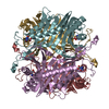
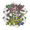
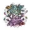
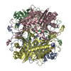
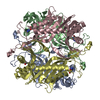
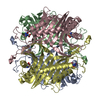
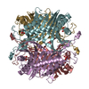
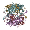
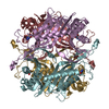
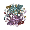
- PDB-6oe8: The crystal structure of hyper-thermostable AgUricase mutant K12C... -
+ Open data

- Basic information
| Entry | Database: PDB / ID: 6oe8 | ||||||
|---|---|---|---|---|---|---|---|
| Title | The crystal structure of hyper-thermostable AgUricase mutant K12C/E286C | ||||||
 Components Components | Uricase | ||||||
 Keywords Keywords | OXIDOREDUCTASE / Uricase / Arthrobacter globiformis / thermostability / disulfide cross-linking | ||||||
| Function / homology |  Function and homology information Function and homology informationfactor-independent urate hydroxylase / urate oxidase activity / urate catabolic process / purine nucleobase metabolic process Similarity search - Function | ||||||
| Biological species |  Arthrobacter globiformis (bacteria) Arthrobacter globiformis (bacteria) | ||||||
| Method |  X-RAY DIFFRACTION / SYNCHROTRON / MOLECULAR REPLACEMENT / Resolution: 1.99 Å X-RAY DIFFRACTION / SYNCHROTRON / MOLECULAR REPLACEMENT / Resolution: 1.99 Å | ||||||
 Authors Authors | Shi, Y. / Wang, T. / Zhou, X.E. / Liu, Q. / Jiang, Y. / Xu, H.E. | ||||||
| Funding support |  China, 1items China, 1items
| ||||||
 Citation Citation | Journal: Acta Pharmacol.Sin. / Year: 2019 Title: Structure-based design of a hyperthermostable AgUricase for hyperuricemia and gout therapy. Authors: Shi, Y. / Wang, T. / Zhou, X.E. / Liu, Q.F. / Jiang, Y. / Xu, H.E. | ||||||
| History |
|
- Structure visualization
| Structure viewer | Molecule: MolmilJmol/JSmol |
|---|
- Downloads & links
-Download
| PDBx/mmCIF format | 6oe8.cif.gz | 274.2 KB | Display | PDBx/mmCIF format |
|---|---|---|---|---|
| PDB format | pdb6oe8.ent.gz | 218.3 KB | Display | PDB format |
| PDBx/mmJSON format | 6oe8.json.gz | Tree view | PDBx/mmJSON format | |
| Others |  Other downloads Other downloads |
-Validation report
| Arichive directory | https://data.pdbj.org/pub/pdb/validation_reports/oe/6oe8ftp://data.pdbj.org/pub/pdb/validation_reports/oe/6oe8 | HTTPS FTP |
|---|
-Related structure data
| Related structure data |  2yzbS S: Starting model for refinement |
|---|---|
| Similar structure data |
-Links
 PDBj
PDBj
- Assembly
| Deposited unit | 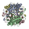
| ||||||||||||
|---|---|---|---|---|---|---|---|---|---|---|---|---|---|
| 1 |
| ||||||||||||
| Unit cell |
| ||||||||||||
| Components on special symmetry positions |
|
-Components
| #1: Protein | Mass: 36611.648 Da / Num. of mol.: 4 / Mutation: K12C, E286C Source method: isolated from a genetically manipulated source Source: (gene. exp.) Arthrobacter globiformis (bacteria) / Gene: uox / Production host: References: UniProt: D0VWQ1, factor-independent urate hydroxylase #2: Chemical | ChemComp-PG4 /   Mass: 194.226 Da / Num. of mol.: 5 / Source method: obtained synthetically / Formula: C8H18O5 / Comment: precipitant*YM Mass: 194.226 Da / Num. of mol.: 5 / Source method: obtained synthetically / Formula: C8H18O5 / Comment: precipitant*YM#3: Chemical | ChemComp-PGE /   Mass: 150.173 Da / Num. of mol.: 8 / Source method: obtained synthetically / Formula: C6H14O4 Mass: 150.173 Da / Num. of mol.: 8 / Source method: obtained synthetically / Formula: C6H14O4#4: Chemical | ChemComp-MLI /   Mass: 102.046 Da / Num. of mol.: 8 / Source method: obtained synthetically / Formula: C3H2O4 Mass: 102.046 Da / Num. of mol.: 8 / Source method: obtained synthetically / Formula: C3H2O4#5: Water | ChemComp-HOH / |  Mass: 18.015 Da / Num. of mol.: 1227 / Source method: isolated from a natural source / Formula: H2O Mass: 18.015 Da / Num. of mol.: 1227 / Source method: isolated from a natural source / Formula: H2OHas protein modification | Y | |
|---|
-Experimental details
-Experiment
| Experiment | Method: X-RAY DIFFRACTION / Number of used crystals: 1 |
|---|
- Sample preparation
| Crystal | Density Matthews: 2.54 Å3/Da / Density % sol: 56.55 % |
|---|---|
| Crystal grow | Temperature: 293 K / Method: vapor diffusion / pH: 5 Details: 12% (w/v) polyethylene glycol 3350 and 0.1M sodium malonate |
-Data collection
| Diffraction | Mean temperature: 100 K / Serial crystal experiment: N |
|---|---|
| Diffraction source | Source: SYNCHROTRON / Site: NFPSS / Beamline: BL19U1 / Wavelength: 1 Å |
| Detector | Type: MARMOSAIC 325 mm CCD / Detector: CCD / Date: Jan 8, 2019 |
| Radiation | Protocol: SINGLE WAVELENGTH / Monochromatic (M) / Laue (L): M / Scattering type: x-ray |
| Radiation wavelength | Wavelength: 1 Å / Relative weight: 1 |
| Reflection | Resolution: 1.99→40 Å / Num. obs: 97576 / % possible obs: 97.4 % / Redundancy: 3.4 % / CC1/2: 0.983 / Rmerge(I) obs: 0.098 / Net I/σ(I): 9.4 |
| Reflection shell | Resolution: 1.99→2.06 Å / Redundancy: 3.3 % / Rmerge(I) obs: 0.308 / Num. unique obs: 9744 / CC1/2: 0.922 / % possible all: 98.3 |
- Processing
| Software |
| |||||||||||||||||||||||||||||||||||||||||||||||||||||||||||||||||||||||||||||||||||||||||||||||||||||||||
|---|---|---|---|---|---|---|---|---|---|---|---|---|---|---|---|---|---|---|---|---|---|---|---|---|---|---|---|---|---|---|---|---|---|---|---|---|---|---|---|---|---|---|---|---|---|---|---|---|---|---|---|---|---|---|---|---|---|---|---|---|---|---|---|---|---|---|---|---|---|---|---|---|---|---|---|---|---|---|---|---|---|---|---|---|---|---|---|---|---|---|---|---|---|---|---|---|---|---|---|---|---|---|---|---|---|---|
| Refinement | Method to determine structure: MOLECULAR REPLACEMENT Starting model: 2YZB Resolution: 1.99→37.276 Å / SU ML: 0.21 / Cross valid method: FREE R-VALUE / σ(F): 1.37 / Phase error: 21.66 / Stereochemistry target values: ML
| |||||||||||||||||||||||||||||||||||||||||||||||||||||||||||||||||||||||||||||||||||||||||||||||||||||||||
| Solvent computation | Shrinkage radii: 0.8 Å / VDW probe radii: 1.1 Å / Solvent model: FLAT BULK SOLVENT MODEL | |||||||||||||||||||||||||||||||||||||||||||||||||||||||||||||||||||||||||||||||||||||||||||||||||||||||||
| Refinement step | Cycle: LAST / Resolution: 1.99→37.276 Å
| |||||||||||||||||||||||||||||||||||||||||||||||||||||||||||||||||||||||||||||||||||||||||||||||||||||||||
| Refine LS restraints |
| |||||||||||||||||||||||||||||||||||||||||||||||||||||||||||||||||||||||||||||||||||||||||||||||||||||||||
| LS refinement shell |
|
