 Movie
Movie Controller

[English] 日本語
 Yorodumi
- PDB-1vay: Crystal Structure of Uricase from Arthrobacter globiformis with i... -
+ Open data

- Basic information
| Entry | Database: PDB / ID: 1vay | ||||||
|---|---|---|---|---|---|---|---|
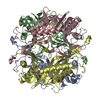
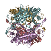
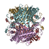
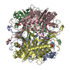
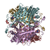
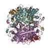
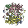
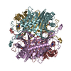
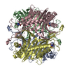
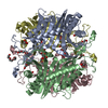
| Title | Crystal Structure of Uricase from Arthrobacter globiformis with inhibitor 8-azaxanthine | ||||||
 Components Components | Uric acid oxidase | ||||||
 Keywords Keywords | OXIDOREDUCTASE | ||||||
| Function / homology | Urate Oxidase / Urate Oxidase; / Roll / Alpha Beta / 8-AZAXANTHINE Function and homology information Function and homology information | ||||||
| Biological species |  Arthrobacter globiformis (bacteria) Arthrobacter globiformis (bacteria) | ||||||
| Method |  X-RAY DIFFRACTION / SYNCHROTRON / MOLECULAR REPLACEMENT / Resolution: 2.24 Å X-RAY DIFFRACTION / SYNCHROTRON / MOLECULAR REPLACEMENT / Resolution: 2.24 Å | ||||||
 Authors Authors | Hossain, M.T. / Suzuki, K. / Yamamoto, T. / Imamura, S. / Sekiguchi, T. / Takenaka, A. | ||||||
 Citation Citation | Journal: To be Published Title: Crystal Structure of Uricase from Arthrobacter Globiformis Complexed with an inhibitor 8-Azaxanthine Authors: Hossain, M.T. / Juan, E.C.M. / Suzuki, K. / Yamamoto, T. / Imamura, S. / Sekiguchi, T. / Takenaka, A. | ||||||
| History |
|
- Structure visualization
| Structure viewer | Molecule: MolmilJmol/JSmol |
|---|
- Downloads & links
-Download
| PDBx/mmCIF format | 1vay.cif.gz | 468.5 KB | Display | PDBx/mmCIF format |
|---|---|---|---|---|
| PDB format | pdb1vay.ent.gz | 386.4 KB | Display | PDB format |
| PDBx/mmJSON format | 1vay.json.gz | Tree view | PDBx/mmJSON format | |
| Others |  Other downloads Other downloads |
-Validation report
| Arichive directory | https://data.pdbj.org/pub/pdb/validation_reports/va/1vayftp://data.pdbj.org/pub/pdb/validation_reports/va/1vay | HTTPS FTP |
|---|
-Related structure data
| Related structure data |  1uox S: Starting model for refinement |
|---|---|
| Similar structure data |


-Links
 PDBj
PDBj- Assembly
| Deposited unit | 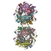
| ||||||||
|---|---|---|---|---|---|---|---|---|---|
| 1 | 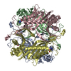
| ||||||||
| 2 | 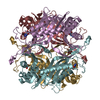
| ||||||||
| Unit cell |
| ||||||||
| Details | The biological assembly is a tetramer |
-Components
| #1: Protein | Mass: 32458.027 Da / Num. of mol.: 8 / Source method: isolated from a natural source / Source: (natural) Arthrobacter globiformis (bacteria) / References: factor-independent urate hydroxylase#2: Chemical | ChemComp-AZA /   Mass: 153.099 Da / Num. of mol.: 8 / Source method: obtained synthetically / Formula: C4H3N5O2 Mass: 153.099 Da / Num. of mol.: 8 / Source method: obtained synthetically / Formula: C4H3N5O2#3: Water | ChemComp-HOH / |  Mass: 18.015 Da / Num. of mol.: 1066 / Source method: isolated from a natural source / Formula: H2O Mass: 18.015 Da / Num. of mol.: 1066 / Source method: isolated from a natural source / Formula: H2O |
|---|
-Experimental details
-Experiment
| Experiment | Method: X-RAY DIFFRACTION / Number of used crystals: 1 |
|---|
- Sample preparation
| Crystal | Density Matthews: 2.79 Å3/Da / Density % sol: 55.84 % |
|---|---|
| Crystal grow | Temperature: 277 K / Method: vapor diffusion / pH: 9 Details: PEG 8000, Lithium sulphate, pH 9.0, VAPOR DIFFUSION, temperature 277K |
-Data collection
| Diffraction | Mean temperature: 100 K |
|---|---|
| Diffraction source | Source: SYNCHROTRON / Site: Photon Factory  / Beamline: AR-NW12A / Wavelength: 1 Å / Beamline: AR-NW12A / Wavelength: 1 Å |
| Detector | Type: ADSC QUANTUM 210 / Detector: CCD / Date: Dec 13, 2003 |
| Radiation | Protocol: SINGLE WAVELENGTH / Monochromatic (M) / Laue (L): M / Scattering type: x-ray |
| Radiation wavelength | Wavelength: 1 Å / Relative weight: 1 |
| Reflection | Resolution: 2.24→20 Å / Num. all: 739628 / Num. obs: 132537 / % possible obs: 97.4 % / Redundancy: 5.4 % / Rmerge(I) obs: 0.067 |
| Reflection shell | Resolution: 2.24→2.32 Å / Redundancy: 5.4 % / Rmerge(I) obs: 0.237 / % possible all: 87.7 |
- Processing
| Software |
| ||||||||||||||||
|---|---|---|---|---|---|---|---|---|---|---|---|---|---|---|---|---|---|
| Refinement | Method to determine structure: MOLECULAR REPLACEMENT Starting model: PDB ENTRY 1UOX 1uox Resolution: 2.24→20 Å / Cross valid method: THROUGHOUT / σ(F): 3
| ||||||||||||||||
| Displacement parameters |
| ||||||||||||||||
| Refine analyze | Luzzati coordinate error obs: 0.23 Å / Luzzati d res low obs: 5 Å | ||||||||||||||||
| Refinement step | Cycle: LAST / Resolution: 2.24→20 Å
| ||||||||||||||||
| Refine LS restraints |
| ||||||||||||||||
| LS refinement shell | Resolution: 2.24→2.32 Å / Rfactor Rfree error: 0.009
|
