 Movie
Movie Controller

[English] 日本語
 Yorodumi
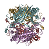
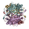
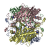
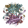
- PDB-2yzd: Crystal structure of uricase from Arthrobacter globiformis in com... -
+ Open data

- Basic information
| Entry | Database: PDB / ID: 2yzd | ||||||
|---|---|---|---|---|---|---|---|
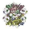
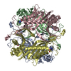
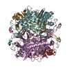
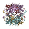
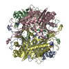
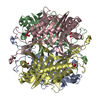
| Title | Crystal structure of uricase from Arthrobacter globiformis in complex with 8-azaxanthin (inhibitor) | ||||||
 Components Components | Uricase | ||||||
 Keywords Keywords | OXIDOREDUCTASE / uricase / 8-azaxanthin | ||||||
| Function / homology |  Function and homology information Function and homology informationfactor-independent urate hydroxylase / urate oxidase activity / urate catabolic process / purine nucleobase metabolic process Similarity search - Function | ||||||
| Biological species |  Arthrobacter globiformis (bacteria) Arthrobacter globiformis (bacteria) | ||||||
| Method |  X-RAY DIFFRACTION / SYNCHROTRON / MOLECULAR REPLACEMENT / Resolution: 2.24 Å X-RAY DIFFRACTION / SYNCHROTRON / MOLECULAR REPLACEMENT / Resolution: 2.24 Å | ||||||
 Authors Authors | Juan, E.C.M. / Hossain, M.T. / Hoque, M.M. / Suzuki, K. / Sekiguchi, T. / Takenaka, A. | ||||||
 Citation Citation | Journal: Acta Crystallogr.,Sect.D / Year: 2008 Title: Structures of Arthrobacter globiformis urate oxidase-ligand complexes. Authors: Juan, E.C. / Hoque, M.M. / Shimizu, S. / Hossain, M.T. / Yamamoto, T. / Imamura, S. / Suzuki, K. / Tsunoda, M. / Amano, H. / Sekiguchi, T. / Takenaka, A. | ||||||
| History |
|
- Structure visualization
| Structure viewer | Molecule: MolmilJmol/JSmol |
|---|
- Downloads & links
-Download
| PDBx/mmCIF format | 2yzd.cif.gz | 470.6 KB | Display | PDBx/mmCIF format |
|---|---|---|---|---|
| PDB format | pdb2yzd.ent.gz | 388 KB | Display | PDB format |
| PDBx/mmJSON format | 2yzd.json.gz | Tree view | PDBx/mmJSON format | |
| Others |  Other downloads Other downloads |
-Validation report
| Arichive directory | https://data.pdbj.org/pub/pdb/validation_reports/yz/2yzdftp://data.pdbj.org/pub/pdb/validation_reports/yz/2yzd | HTTPS FTP |
|---|
-Related structure data
| Related structure data |  2yzbC  2yzcC  2yzeC  1uox C: citing same article ( S: Starting model for refinement |
|---|---|
| Similar structure data |


-Links
 PDBj
PDBj
- Assembly
| Deposited unit | 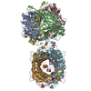
| ||||||||
|---|---|---|---|---|---|---|---|---|---|
| 1 | 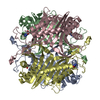
| ||||||||
| 2 | 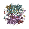
| ||||||||
| Unit cell |
|
-Components
| #1: Protein | Mass: 33900.629 Da / Num. of mol.: 8 / Source method: isolated from a natural source / Source: (natural) Arthrobacter globiformis (bacteria)References: UniProt: D0VWQ1*PLUS, factor-independent urate hydroxylase #2: Chemical | ChemComp-AZA / 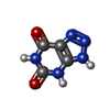  Mass: 153.099 Da / Num. of mol.: 8 / Source method: obtained synthetically / Formula: C4H3N5O2 Mass: 153.099 Da / Num. of mol.: 8 / Source method: obtained synthetically / Formula: C4H3N5O2#3: Water | ChemComp-HOH / |  Mass: 18.015 Da / Num. of mol.: 1160 / Source method: isolated from a natural source / Formula: H2O Mass: 18.015 Da / Num. of mol.: 1160 / Source method: isolated from a natural source / Formula: H2OHas protein modification | N | Sequence details | THE SEQUENCE DATABASE FOR THIS PROTEIN DOES NOT CURRENTLY EXIST IN UNP. IT WILL BE SUBMITTED TO UNP ...THE SEQUENCE DATABASE FOR THIS PROTEIN DOES NOT CURRENTLY EXIST IN UNP. IT WILL BE SUBMITTED TO UNP DATABASE SOON. | |
|---|
-Experimental details
-Experiment
| Experiment | Method: X-RAY DIFFRACTION / Number of used crystals: 1 |
|---|
- Sample preparation
| Crystal | Density Matthews: 2.67 Å3/Da / Density % sol: 53.87 % |
|---|---|
| Crystal grow | Temperature: 277 K / Method: vapor diffusion, hanging drop / pH: 9 Details: PEG 8000, lithium sulfate, sodium borate buffer, pH 9.0, VAPOR DIFFUSION, HANGING DROP, temperature 277K |
-Data collection
| Diffraction | Mean temperature: 100 K |
|---|---|
| Diffraction source | Source: SYNCHROTRON / Site: Photon Factory  / Beamline: AR-NW12A / Wavelength: 1 Å / Beamline: AR-NW12A / Wavelength: 1 Å |
| Detector | Type: ADSC QUANTUM 210 / Detector: CCD / Date: Dec 12, 2003 |
| Radiation | Monochromator: SI(111) / Protocol: SINGLE WAVELENGTH / Monochromatic (M) / Laue (L): M / Scattering type: x-ray |
| Radiation wavelength | Wavelength: 1 Å / Relative weight: 1 |
| Reflection | Resolution: 2.24→20 Å / Num. obs: 132537 / % possible obs: 97.4 % / Observed criterion σ(F): 0 / Observed criterion σ(I): 0 / Redundancy: 5.4 % / Rmerge(I) obs: 0.067 |
| Reflection shell | Resolution: 2.24→2.32 Å / Redundancy: 5.4 % / Rmerge(I) obs: 0.237 / % possible all: 87.7 |
- Processing
| Software |
| |||||||||||||||||||||||||
|---|---|---|---|---|---|---|---|---|---|---|---|---|---|---|---|---|---|---|---|---|---|---|---|---|---|---|
| Refinement | Method to determine structure: MOLECULAR REPLACEMENT Starting model: PDB entry 1uox 1uox Resolution: 2.24→20 Å / Cross valid method: THROUGHOUT / σ(F): 3
| |||||||||||||||||||||||||
| Displacement parameters |
| |||||||||||||||||||||||||
| Refinement step | Cycle: LAST / Resolution: 2.24→20 Å
|
