 Movie
Movie Controller

[English] 日本語
 Yorodumi
- PDB-6oad: 2.05 Angstrom Resolution Crystal Structure of Aminopeptidase B fr... -
+ Open data

- Basic information
| Entry | Database: PDB / ID: 6oad | ||||||
|---|---|---|---|---|---|---|---|
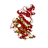
| Title | 2.05 Angstrom Resolution Crystal Structure of Aminopeptidase B from Escherichia coli str. K-12 substr. MG1655. | ||||||
 Components Components | Peptidase B | ||||||
 Keywords Keywords | HYDROLASE / Structural Genomics / Center for Structural Genomics of Infectious Diseases / CSGID / Aminopeptidase B | ||||||
| Function / homology |  Function and homology information Function and homology informationPepB aminopeptidase / peptide catabolic process / metalloaminopeptidase activity / aminopeptidase activity / peptidase activity / manganese ion binding / proteolysis / identical protein binding / cytoplasm / cytosol Similarity search - Function | ||||||
| Biological species |  | ||||||
| Method |  X-RAY DIFFRACTION / SYNCHROTRON / MOLECULAR REPLACEMENT / Resolution: 2.05 Å X-RAY DIFFRACTION / SYNCHROTRON / MOLECULAR REPLACEMENT / Resolution: 2.05 Å | ||||||
 Authors Authors | Minasov, G. / Shuvalova, L. / Wawrzak, Z. / Kiryukhina, O. / Grimshaw, S. / Kwon, K. / Satchell, K.J.F. / Center for Structural Genomics of Infectious Diseases (CSGID) | ||||||
 Citation Citation | Journal: Protein Sci. / Year: 2020 Title: Comparison of metal-bound and unbound structures of aminopeptidase B proteins from Escherichia coli and Yersinia pestis. Authors: Minasov, G. / Lam, M.R. / Rosas-Lemus, M. / Slawek, J. / Woinska, M. / Shabalin, I.G. / Shuvalova, L. / Palsson, B.O. / Godzik, A. / Minor, W. / Satchell, K.J.F. #1: Journal: Acta Crystallogr.,Sect.F / Year: 2019Title: Structural comparison of p-hydroxybenzoate hydroxylase (PobA) from Pseudomonas putida with PobA from other Pseudomonas spp. and other monooxygenases. Authors: Lazar, J.T. / Shuvalova, L. / Rosas-Lemus, M. / Kiryukhina, O. / Satchell, K.J.F. / Minasov, G. #2: Journal: Febs J. / Year: 2020Title: Structural and biochemical analysis of Bacillus anthracis prephenate dehydrogenase reveals an unusual mode of inhibition by tyrosine via the ACT domain. Authors: Shabalin, I.G. / Gritsunov, A. / Hou, J. / Slawek, J. / Miks, C.D. / Cooper, D.R. / Minor, W. / Christendat, D. | ||||||
| History |
|
- Structure visualization
| Structure viewer | Molecule: MolmilJmol/JSmol |
|---|
- Downloads & links
-Download
| PDBx/mmCIF format | 6oad.cif.gz | 1.9 MB | Display | PDBx/mmCIF format |
|---|---|---|---|---|
| PDB format | pdb6oad.ent.gz | 1.6 MB | Display | PDB format |
| PDBx/mmJSON format | 6oad.json.gz | Tree view | PDBx/mmJSON format | |
| Others |  Other downloads Other downloads |
-Validation report
| Arichive directory | https://data.pdbj.org/pub/pdb/validation_reports/oa/6oadftp://data.pdbj.org/pub/pdb/validation_reports/oa/6oad | HTTPS FTP |
|---|
-Related structure data
| Related structure data |  6ov8C  3ij3S S: Starting model for refinement C: citing same article ( |
|---|---|
| Similar structure data | |
| Other databases |


-Links
 PDBj
PDBj


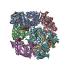
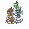
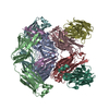
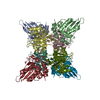
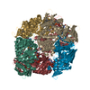
- Assembly
| Deposited unit | 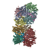
| ||||||||
|---|---|---|---|---|---|---|---|---|---|
| 1 | 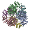
| ||||||||
| 2 | 
| ||||||||
| Unit cell |
|
-Components
-Protein , 1 types, 12 molecules ABCDEFGHIJKL
| #1: Protein | Mass: 46502.270 Da / Num. of mol.: 12 Source method: isolated from a genetically manipulated source Source: (gene. exp.) Gene: pepB, D8B36_06045 / Variant: MG1655 / Plasmid: pMCSG53 / Production host: References: UniProt: A0A387CSU7, UniProt: P37095*PLUS, PepB aminopeptidase |
|---|
-Non-polymers , 8 types, 3495 molecules 


| #2: Chemical | ChemComp-ZN /  Mass: 65.409 Da / Num. of mol.: 24 / Source method: obtained synthetically / Formula: Zn Mass: 65.409 Da / Num. of mol.: 24 / Source method: obtained synthetically / Formula: Zn#3: Chemical | ChemComp-CA /  Mass: 40.078 Da / Num. of mol.: 13 / Source method: obtained synthetically / Formula: Ca Mass: 40.078 Da / Num. of mol.: 13 / Source method: obtained synthetically / Formula: Ca#4: Chemical | ChemComp-CL /  Mass: 35.453 Da / Num. of mol.: 9 / Source method: obtained synthetically / Formula: Cl Mass: 35.453 Da / Num. of mol.: 9 / Source method: obtained synthetically / Formula: Cl#5: Chemical | ChemComp-BCT /  Mass: 61.017 Da / Num. of mol.: 19 / Source method: obtained synthetically / Formula: CHO3 / Comment: pH buffer*YM Mass: 61.017 Da / Num. of mol.: 19 / Source method: obtained synthetically / Formula: CHO3 / Comment: pH buffer*YM#6: Chemical | ChemComp-EDO /  Mass: 62.068 Da / Num. of mol.: 39 / Source method: obtained synthetically / Formula: C2H6O2 Mass: 62.068 Da / Num. of mol.: 39 / Source method: obtained synthetically / Formula: C2H6O2#7: Chemical | ChemComp-PGE / |  Mass: 150.173 Da / Num. of mol.: 1 / Source method: obtained synthetically / Formula: C6H14O4 Mass: 150.173 Da / Num. of mol.: 1 / Source method: obtained synthetically / Formula: C6H14O4#8: Chemical | ChemComp-PEG /  Mass: 106.120 Da / Num. of mol.: 5 / Source method: obtained synthetically / Formula: C4H10O3 Mass: 106.120 Da / Num. of mol.: 5 / Source method: obtained synthetically / Formula: C4H10O3#9: Water | ChemComp-HOH / | Mass: 18.015 Da / Num. of mol.: 3385 / Source method: isolated from a natural source / Formula: H2O |
|---|
-Experimental details
-Experiment
| Experiment | Method: X-RAY DIFFRACTION / Number of used crystals: 1 |
|---|
- Sample preparation
| Crystal | Density Matthews: 2.4 Å3/Da / Density % sol: 48.8 % |
|---|---|
| Crystal grow | Temperature: 292 K / Method: vapor diffusion, sitting drop / pH: 7.5 Details: Protein: 6.0 mg/ml, 0.01M Tris pH 8.3; Screen: PEGs II (H8), 0.2M Calcium acetate, 0.1M HEPES pH 7.5, 10% (w/v) PEG 8000. Cryo: reservoir + 20% Ethylene glycol. |
-Data collection
| Diffraction | Mean temperature: 100 K / Serial crystal experiment: N |
|---|---|
| Diffraction source | Source: SYNCHROTRON / Site: APS  / Beamline: 21-ID-F / Wavelength: 0.97872 Å / Beamline: 21-ID-F / Wavelength: 0.97872 Å |
| Detector | Type: MARMOSAIC 300 mm CCD / Detector: CCD / Date: Feb 5, 2019 / Details: C(111) |
| Radiation | Monochromator: Be / Protocol: SINGLE WAVELENGTH / Monochromatic (M) / Laue (L): M / Scattering type: x-ray |
| Radiation wavelength | Wavelength: 0.97872 Å / Relative weight: 1 |
| Reflection | Resolution: 2.05→30 Å / Num. obs: 338775 / % possible obs: 98.8 % / Observed criterion σ(I): -3 / Redundancy: 4.1 % / Biso Wilson estimate: 30.9 Å2 / Rmerge(I) obs: 0.098 / Rpim(I) all: 0.054 / Rrim(I) all: 0.112 / Rsym value: 0.098 / Χ2: 1.011 / Net I/σ(I): 11.9 |
| Reflection shell | Resolution: 2.05→2.09 Å / Redundancy: 4.1 % / Rmerge(I) obs: 0.791 / Mean I/σ(I) obs: 1.9 / Num. unique obs: 16506 / CC1/2: 0.761 / Rpim(I) all: 0.437 / Rrim(I) all: 0.905 / Rsym value: 0.791 / Χ2: 1.007 / % possible all: 96.4 |
- Processing
| Software |
| ||||||||||||||||||||||||||||||||||||||||||||||||||||||||||||||||||||||||||||||||||||||||||||||||||||||||||||||||||||||||||||||||||||||||||||||||||||||||||||||||||||||||||||||||||||||
|---|---|---|---|---|---|---|---|---|---|---|---|---|---|---|---|---|---|---|---|---|---|---|---|---|---|---|---|---|---|---|---|---|---|---|---|---|---|---|---|---|---|---|---|---|---|---|---|---|---|---|---|---|---|---|---|---|---|---|---|---|---|---|---|---|---|---|---|---|---|---|---|---|---|---|---|---|---|---|---|---|---|---|---|---|---|---|---|---|---|---|---|---|---|---|---|---|---|---|---|---|---|---|---|---|---|---|---|---|---|---|---|---|---|---|---|---|---|---|---|---|---|---|---|---|---|---|---|---|---|---|---|---|---|---|---|---|---|---|---|---|---|---|---|---|---|---|---|---|---|---|---|---|---|---|---|---|---|---|---|---|---|---|---|---|---|---|---|---|---|---|---|---|---|---|---|---|---|---|---|---|---|---|---|
| Refinement | Method to determine structure: MOLECULAR REPLACEMENT Starting model: 3IJ3 Resolution: 2.05→29.88 Å / Cor.coef. Fo:Fc: 0.968 / Cor.coef. Fo:Fc free: 0.948 / SU B: 11.004 / SU ML: 0.152 / Cross valid method: THROUGHOUT / ESU R: 0.194 / ESU R Free: 0.165 / Details: HYDROGENS HAVE BEEN ADDED IN THE RIDING POSITIONS
| ||||||||||||||||||||||||||||||||||||||||||||||||||||||||||||||||||||||||||||||||||||||||||||||||||||||||||||||||||||||||||||||||||||||||||||||||||||||||||||||||||||||||||||||||||||||
| Solvent computation | Ion probe radii: 0.8 Å / Shrinkage radii: 0.8 Å / VDW probe radii: 1.2 Å | ||||||||||||||||||||||||||||||||||||||||||||||||||||||||||||||||||||||||||||||||||||||||||||||||||||||||||||||||||||||||||||||||||||||||||||||||||||||||||||||||||||||||||||||||||||||
| Displacement parameters | Biso mean: 37.118 Å2
| ||||||||||||||||||||||||||||||||||||||||||||||||||||||||||||||||||||||||||||||||||||||||||||||||||||||||||||||||||||||||||||||||||||||||||||||||||||||||||||||||||||||||||||||||||||||
| Refinement step | Cycle: 1 / Resolution: 2.05→29.88 Å
| ||||||||||||||||||||||||||||||||||||||||||||||||||||||||||||||||||||||||||||||||||||||||||||||||||||||||||||||||||||||||||||||||||||||||||||||||||||||||||||||||||||||||||||||||||||||
| Refine LS restraints |
|