 Movie
Movie Controller

[English] 日本語
 Yorodumi
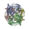
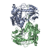
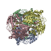
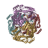
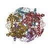
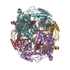
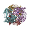
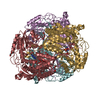
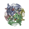
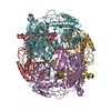
- PDB-6o4i: Structure of ALDH7A1 mutant E399D complexed with alpha-aminoadipate -
+ Open data

- Basic information
| Entry | Database: PDB / ID: 6o4i | |||||||||
|---|---|---|---|---|---|---|---|---|---|---|
| Title | Structure of ALDH7A1 mutant E399D complexed with alpha-aminoadipate | |||||||||
 Components Components | Alpha-aminoadipic semialdehyde dehydrogenase | |||||||||
 Keywords Keywords | OXIDOREDUCTASE / ALDEHYDE DEHYDROGENASE / NAD / LYSINE CATABOLISM | |||||||||
| Function / homology |  Function and homology information Function and homology informationnegative regulation of endosome to plasma membrane protein transport / L-aminoadipate-semialdehyde dehydrogenase [NAD(P)+] activity / L-aminoadipate-semialdehyde dehydrogenase / Choline catabolism / negative regulation of Golgi to plasma membrane protein transport / L-lysine catabolic process / choline catabolic process / Lysine catabolism / : / betaine-aldehyde dehydrogenase (NAD+) activity ...negative regulation of endosome to plasma membrane protein transport / L-aminoadipate-semialdehyde dehydrogenase [NAD(P)+] activity / L-aminoadipate-semialdehyde dehydrogenase / Choline catabolism / negative regulation of Golgi to plasma membrane protein transport / L-lysine catabolic process / choline catabolic process / Lysine catabolism / : / betaine-aldehyde dehydrogenase (NAD+) activity / betaine-aldehyde dehydrogenase / : / aldehyde metabolic process / plasma membrane to endosome transport / aldehyde dehydrogenase (NAD+) / Golgi to endosome transport / aldehyde dehydrogenase (NAD+) activity / oxidoreductase activity, acting on NAD(P)H, quinone or similar compound as acceptor / endosome to lysosome transport / negative regulation of ferroptosis / energy homeostasis / sensory perception of sound / mitochondrial matrix / Golgi membrane / mitochondrion / extracellular exosome / identical protein binding / nucleus / plasma membrane / cytosol Similarity search - Function | |||||||||
| Biological species |  Homo sapiens (human) Homo sapiens (human) | |||||||||
| Method |  X-RAY DIFFRACTION / SYNCHROTRON / FOURIER SYNTHESIS / Resolution: 1.75 Å X-RAY DIFFRACTION / SYNCHROTRON / FOURIER SYNTHESIS / Resolution: 1.75 Å | |||||||||
 Authors Authors | Tanner, J.J. / Korasick, D.A. / Laciak, A.R. | |||||||||
| Funding support |  United States, 1items United States, 1items
| |||||||||
 Citation Citation | Journal: J Inherit Metab Dis / Year: 2020 Title: Structural analysis of pathogenic mutations targeting Glu427 of ALDH7A1, the hot spot residue of pyridoxine-dependent epilepsy. Authors: Adrian R Laciak / David A Korasick / Kent S Gates / John J Tanner / Abstract: Certain loss-of-function mutations in the gene encoding the lysine catabolic enzyme aldehyde dehydrogenase 7A1 (ALDH7A1) cause pyridoxine-dependent epilepsy (PDE). Missense mutations of Glu427, ...Certain loss-of-function mutations in the gene encoding the lysine catabolic enzyme aldehyde dehydrogenase 7A1 (ALDH7A1) cause pyridoxine-dependent epilepsy (PDE). Missense mutations of Glu427, especially Glu427Gln, account for ~30% of the mutated alleles in PDE patients, and thus Glu427 has been referred to as a mutation hot spot of PDE. Glu427 is invariant in the ALDH superfamily and forms ionic hydrogen bonds with the nicotinamide ribose of the NAD cofactor. Here we report the first crystal structures of ALDH7A1 containing pathogenic mutations targeting Glu427. The mutant enzymes E427Q, Glu427Asp, and Glu427Gly were expressed in Escherichia coli and purified. The recombinant enzymes displayed negligible catalytic activity compared to the wild-type enzyme. The crystal structures of the mutant enzymes complexed with NAD were determined to understand how the mutations impact NAD binding. In the E427Q and E427G structures, the nicotinamide mononucleotide is highly flexible and lacks a defined binding pose. In E427D, the bound NAD adopts a "retracted" conformation in which the nicotinamide ring is too far from the catalytic Cys residue for hydride transfer. Thus, the structures revealed a shared mechanism for loss of function: none of the variants are able to stabilise the nicotinamide of NAD in the pose required for catalysis. We also show that these mutations reduce the amount of active tetrameric ALDH7A1 at the concentration of NAD tested. Altogether, our results provide the three-dimensional molecular structural basis of the most common pathogenic variants of PDE and implicate strong (ionic) hydrogen bonds in the aetiology of a human disease. | |||||||||
| History |
|
- Structure visualization
| Structure viewer | Molecule: MolmilJmol/JSmol |
|---|
- Downloads & links
-Download
| PDBx/mmCIF format | 6o4i.cif.gz | 1.5 MB | Display | PDBx/mmCIF format |
|---|---|---|---|---|
| PDB format | pdb6o4i.ent.gz | 1.3 MB | Display | PDB format |
| PDBx/mmJSON format | 6o4i.json.gz | Tree view | PDBx/mmJSON format | |
| Others |  Other downloads Other downloads |
-Validation report
| Arichive directory | https://data.pdbj.org/pub/pdb/validation_reports/o4/6o4iftp://data.pdbj.org/pub/pdb/validation_reports/o4/6o4i | HTTPS FTP |
|---|
-Related structure data
| Related structure data | 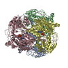 6o4kC 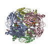 6o4lC 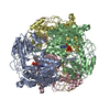 6u2xC  4zulS S: Starting model for refinement C: citing same article ( |
|---|---|
| Similar structure data |


-Links
 PDBj
PDBj

- Assembly
| Deposited unit | 
| ||||||||
|---|---|---|---|---|---|---|---|---|---|
| 1 | 
| ||||||||
| 2 | 
| ||||||||
| Unit cell |
|
-Components
| #1: Protein | Mass: 55606.340 Da / Num. of mol.: 8 / Mutation: E399D Source method: isolated from a genetically manipulated source Source: (gene. exp.) Homo sapiens (human) / Gene: ALDH7A1, ATQ1 / Production host:  References: UniProt: P49419, L-aminoadipate-semialdehyde dehydrogenase, aldehyde dehydrogenase (NAD+), betaine-aldehyde dehydrogenase #2: Chemical | ChemComp-UN1 / 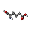  Type: L-peptide linking / Mass: 161.156 Da / Num. of mol.: 8 / Source method: obtained synthetically / Formula: C6H11NO4 / Feature type: SUBJECT OF INVESTIGATION Type: L-peptide linking / Mass: 161.156 Da / Num. of mol.: 8 / Source method: obtained synthetically / Formula: C6H11NO4 / Feature type: SUBJECT OF INVESTIGATION#3: Chemical | ChemComp-EDO /   Mass: 62.068 Da / Num. of mol.: 16 / Source method: obtained synthetically / Formula: C2H6O2 Mass: 62.068 Da / Num. of mol.: 16 / Source method: obtained synthetically / Formula: C2H6O2#4: Water | ChemComp-HOH / |  Mass: 18.015 Da / Num. of mol.: 1925 / Source method: isolated from a natural source / Formula: H2O Mass: 18.015 Da / Num. of mol.: 1925 / Source method: isolated from a natural source / Formula: H2O |
|---|
-Experimental details
-Experiment
| Experiment | Method: X-RAY DIFFRACTION / Number of used crystals: 1 |
|---|
- Sample preparation
| Crystal | Density Matthews: 2.2 Å3/Da / Density % sol: 43.97 % |
|---|---|
| Crystal grow | Temperature: 293 K / Method: vapor diffusion, sitting drop / pH: 5.4 Details: 0.2 M MgCl2, 0.1 M Bis-Tris pH 5.4, and 25% (w/v) PEG 3350 |
-Data collection
| Diffraction | Mean temperature: 100 K / Serial crystal experiment: N | ||||||||||||||||||||||||
|---|---|---|---|---|---|---|---|---|---|---|---|---|---|---|---|---|---|---|---|---|---|---|---|---|---|
| Diffraction source | Source: SYNCHROTRON / Site: ALS / Beamline: 4.2.2 / Wavelength: 1 Å | ||||||||||||||||||||||||
| Detector | Type: RDI CMOS_8M / Detector: CMOS / Date: Oct 4, 2018 | ||||||||||||||||||||||||
| Radiation | Protocol: SINGLE WAVELENGTH / Monochromatic (M) / Laue (L): M / Scattering type: x-ray | ||||||||||||||||||||||||
| Radiation wavelength | Wavelength: 1 Å / Relative weight: 1 | ||||||||||||||||||||||||
| Reflection | Resolution: 1.75→49.03 Å / Num. obs: 379396 / % possible obs: 98.6 % / Redundancy: 8.2 % / CC1/2: 0.998 / Rmerge(I) obs: 0.093 / Rpim(I) all: 0.031 / Rrim(I) all: 0.098 / Net I/σ(I): 12.2 / Num. measured all: 3102032 / Scaling rejects: 363 | ||||||||||||||||||||||||
| Reflection shell | Diffraction-ID: 1
|
- Processing
| Software |
| |||||||||||||||||||||||||||||||||||||||||||||||||||||||||||||||||||||||||||||||||||||||||||||||||||||||||||||||||||||||||||||||||||||||||||||||||||||||||||||||||||||||||||||||||||||||||||||||||||||||||||||||||||||||||||||||||
|---|---|---|---|---|---|---|---|---|---|---|---|---|---|---|---|---|---|---|---|---|---|---|---|---|---|---|---|---|---|---|---|---|---|---|---|---|---|---|---|---|---|---|---|---|---|---|---|---|---|---|---|---|---|---|---|---|---|---|---|---|---|---|---|---|---|---|---|---|---|---|---|---|---|---|---|---|---|---|---|---|---|---|---|---|---|---|---|---|---|---|---|---|---|---|---|---|---|---|---|---|---|---|---|---|---|---|---|---|---|---|---|---|---|---|---|---|---|---|---|---|---|---|---|---|---|---|---|---|---|---|---|---|---|---|---|---|---|---|---|---|---|---|---|---|---|---|---|---|---|---|---|---|---|---|---|---|---|---|---|---|---|---|---|---|---|---|---|---|---|---|---|---|---|---|---|---|---|---|---|---|---|---|---|---|---|---|---|---|---|---|---|---|---|---|---|---|---|---|---|---|---|---|---|---|---|---|---|---|---|---|---|---|---|---|---|---|---|---|---|---|---|---|---|---|---|---|
| Refinement | Method to determine structure: FOURIER SYNTHESIS Starting model: 4zul Resolution: 1.75→49.028 Å / SU ML: 0.28 / Cross valid method: THROUGHOUT / σ(F): 0.31 / Phase error: 30.04
| |||||||||||||||||||||||||||||||||||||||||||||||||||||||||||||||||||||||||||||||||||||||||||||||||||||||||||||||||||||||||||||||||||||||||||||||||||||||||||||||||||||||||||||||||||||||||||||||||||||||||||||||||||||||||||||||||
| Solvent computation | Shrinkage radii: 0.9 Å / VDW probe radii: 1.11 Å | |||||||||||||||||||||||||||||||||||||||||||||||||||||||||||||||||||||||||||||||||||||||||||||||||||||||||||||||||||||||||||||||||||||||||||||||||||||||||||||||||||||||||||||||||||||||||||||||||||||||||||||||||||||||||||||||||
| Displacement parameters | Biso max: 100.61 Å2 / Biso mean: 38.6801 Å2 / Biso min: 14.3 Å2 | |||||||||||||||||||||||||||||||||||||||||||||||||||||||||||||||||||||||||||||||||||||||||||||||||||||||||||||||||||||||||||||||||||||||||||||||||||||||||||||||||||||||||||||||||||||||||||||||||||||||||||||||||||||||||||||||||
| Refinement step | Cycle: final / Resolution: 1.75→49.028 Å
| |||||||||||||||||||||||||||||||||||||||||||||||||||||||||||||||||||||||||||||||||||||||||||||||||||||||||||||||||||||||||||||||||||||||||||||||||||||||||||||||||||||||||||||||||||||||||||||||||||||||||||||||||||||||||||||||||
| LS refinement shell | Refine-ID: X-RAY DIFFRACTION / Rfactor Rfree error: 0 / Total num. of bins used: 30
| |||||||||||||||||||||||||||||||||||||||||||||||||||||||||||||||||||||||||||||||||||||||||||||||||||||||||||||||||||||||||||||||||||||||||||||||||||||||||||||||||||||||||||||||||||||||||||||||||||||||||||||||||||||||||||||||||
| Refinement TLS params. | Method: refined / Refine-ID: X-RAY DIFFRACTION
| |||||||||||||||||||||||||||||||||||||||||||||||||||||||||||||||||||||||||||||||||||||||||||||||||||||||||||||||||||||||||||||||||||||||||||||||||||||||||||||||||||||||||||||||||||||||||||||||||||||||||||||||||||||||||||||||||
| Refinement TLS group |
|
