 Movie
Movie Controller
[English] 日本語
 Yorodumi
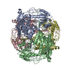
- SASDGQ4: Human alpha-aminoadipic semialdehyde dehydrogenase (ALDH)7A1 E399... -
+ Open data

- Basic information
| Entry | Database: SASBDB / ID: SASDGQ4 |
|---|---|
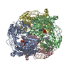
 Sample Sample | Human alpha-aminoadipic semialdehyde dehydrogenase (ALDH)7A1 E399D at 2.9 mg/mL
|
| Function / homology |  Function and homology information Function and homology informationnegative regulation of endosome to plasma membrane protein transport / L-aminoadipate-semialdehyde dehydrogenase [NAD(P)+] activity / L-aminoadipate-semialdehyde dehydrogenase / Choline catabolism / negative regulation of Golgi to plasma membrane protein transport / L-lysine catabolic process / choline catabolic process / Lysine catabolism / : / betaine-aldehyde dehydrogenase (NAD+) activity ...negative regulation of endosome to plasma membrane protein transport / L-aminoadipate-semialdehyde dehydrogenase [NAD(P)+] activity / L-aminoadipate-semialdehyde dehydrogenase / Choline catabolism / negative regulation of Golgi to plasma membrane protein transport / L-lysine catabolic process / choline catabolic process / Lysine catabolism / : / betaine-aldehyde dehydrogenase (NAD+) activity / betaine-aldehyde dehydrogenase / : / aldehyde metabolic process / plasma membrane to endosome transport / aldehyde dehydrogenase (NAD+) / Golgi to endosome transport / aldehyde dehydrogenase (NAD+) activity / oxidoreductase activity, acting on NAD(P)H, quinone or similar compound as acceptor / endosome to lysosome transport / negative regulation of ferroptosis / energy homeostasis / sensory perception of sound / mitochondrial matrix / Golgi membrane / mitochondrion / extracellular exosome / identical protein binding / nucleus / plasma membrane / cytosol Similarity search - Function |
| Biological species |  Homo sapiens (human) Homo sapiens (human) |
 Citation Citation | Journal: J Inherit Metab Dis / Year: 2020 Title: Structural analysis of pathogenic mutations targeting Glu427 of ALDH7A1, the hot spot residue of pyridoxine-dependent epilepsy. Authors: Adrian R Laciak / David A Korasick / Kent S Gates / John J Tanner /  Abstract: Certain loss-of-function mutations in the gene encoding the lysine catabolic enzyme aldehyde dehydrogenase 7A1 (ALDH7A1) cause pyridoxine-dependent epilepsy (PDE). Missense mutations of Glu427, ...Certain loss-of-function mutations in the gene encoding the lysine catabolic enzyme aldehyde dehydrogenase 7A1 (ALDH7A1) cause pyridoxine-dependent epilepsy (PDE). Missense mutations of Glu427, especially Glu427Gln, account for ~30% of the mutated alleles in PDE patients, and thus Glu427 has been referred to as a mutation hot spot of PDE. Glu427 is invariant in the ALDH superfamily and forms ionic hydrogen bonds with the nicotinamide ribose of the NAD cofactor. Here we report the first crystal structures of ALDH7A1 containing pathogenic mutations targeting Glu427. The mutant enzymes E427Q, Glu427Asp, and Glu427Gly were expressed in Escherichia coli and purified. The recombinant enzymes displayed negligible catalytic activity compared to the wild-type enzyme. The crystal structures of the mutant enzymes complexed with NAD were determined to understand how the mutations impact NAD binding. In the E427Q and E427G structures, the nicotinamide mononucleotide is highly flexible and lacks a defined binding pose. In E427D, the bound NAD adopts a "retracted" conformation in which the nicotinamide ring is too far from the catalytic Cys residue for hydride transfer. Thus, the structures revealed a shared mechanism for loss of function: none of the variants are able to stabilise the nicotinamide of NAD in the pose required for catalysis. We also show that these mutations reduce the amount of active tetrameric ALDH7A1 at the concentration of NAD tested. Altogether, our results provide the three-dimensional molecular structural basis of the most common pathogenic variants of PDE and implicate strong (ionic) hydrogen bonds in the aetiology of a human disease. |
 Contact author Contact author |
|
- Structure visualization
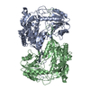
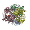
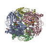
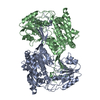
| Structure viewer | Molecule: MolmilJmol/JSmol |
|---|
- Downloads & links
-Data source
| SASBDB page |  SASDGQ4 SASDGQ4 |
|---|
-Related structure data


-External links
| Related items in Molecule of the Month |
|---|


-Models
| Model #3623 |   Type: atomic / Chi-square value: 1.04650799044264  Search similar-shape structures of this assembly by Omokage search (details) Search similar-shape structures of this assembly by Omokage search (details) |
|---|---|
| Model #3624 |  Type: atomic / Chi-square value: 1.04650799044264 Search similar-shape structures of this assembly by Omokage search (details) |
-Sample
| Sample | Name: Human alpha-aminoadipic semialdehyde dehydrogenase (ALDH)7A1 E399D at 2.9 mg/mL Specimen concentration: 2.9 mg/ml |
|---|---|
| Buffer | Name: 50 mM HEPES, 100 mM NaCl, 1 mM DTT, 10 mM NAD, 5% (v/v) glycerol pH: 8 |
| Entity #1846 | Type: protein / Description: Alpha-aminoadipic semialdehyde dehydrogenase / Formula weight: 55.546 / Source: Homo sapiens / References: UniProt: P49419 Sequence: GHMSTLLINQ PQYAWLKELG LREENEGVYN GSWGGRGEVI TTYCPANNEP IARVRQASVA DYEETVKKAR EAWKIWADIP APKRGEIVRQ IGDALREKIQ VLGSLVSLEM GKILVEGVGE VQEYVDICDY AVGLSRMIGG PILPSERSGH ALIEQWNPVG LVGIITAFNF ...Sequence: GHMSTLLINQ PQYAWLKELG LREENEGVYN GSWGGRGEVI TTYCPANNEP IARVRQASVA DYEETVKKAR EAWKIWADIP APKRGEIVRQ IGDALREKIQ VLGSLVSLEM GKILVEGVGE VQEYVDICDY AVGLSRMIGG PILPSERSGH ALIEQWNPVG LVGIITAFNF PVAVYGWNNA IAMICGNVCL WKGAPTTSLI SVAVTKIIAK VLEDNKLPGA ICSLTCGGAD IGTAMAKDER VNLLSFTGST QVGKQVGLMV QERFGRSLLE LGGNNAIIAF EDADLSLVVP SALFAAVGTA GQRCTTARRL FIHESIHDEV VNRLKKAYAQ IRVGNPWDPN VLYGPLHTKQ AVSMFLGAVE EAKKEGGTVV YGGKVMDRPG NYVEPTIVTG LGHDASIAHT DTFAPILYVF KFKNEEEVFA WNNEVKQGLS SSIFTKDLGR IFRWLGPKGS DCGIVNVNIP TSGAEIGGAF GGEKHTGGGR ESGSDAWKQY MRRSTCTINY SKDLPLAQGI KFQ |
-Experimental information
| Beam | Instrument name: Advanced Light Source (ALS) 12.3.1 (SIBYLS) City: Berkeley, CA / 国: USA / Type of source: X-ray synchrotron / Wavelength: 0.127 Å / Dist. spec. to detc.: 2 mm | ||||||||||||||||||||||||||||||
|---|---|---|---|---|---|---|---|---|---|---|---|---|---|---|---|---|---|---|---|---|---|---|---|---|---|---|---|---|---|---|---|
| Detector | Name: Pilatus3 X 2M / Pixsize x: 172 mm | ||||||||||||||||||||||||||||||
| Scan |  Measurement date: May 1, 2019 / Storage temperature: 4 °C / Cell temperature: 10 °C / Unit: 1/A /
| ||||||||||||||||||||||||||||||
| Result | Type of curve: single_conc /
|
