- PDB-6o2d: Schizosaccharomyces pombe Cnp3 Cupin Domain -
+
Open data
ID or keywords:
Loading...
-
Basic information
Entry
Database: PDB / ID: 6o2d
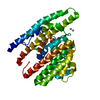
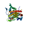
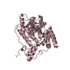
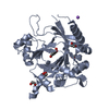
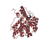
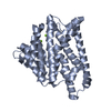
Title
Schizosaccharomyces pombe Cnp3 Cupin Domain
Components
Inner kinetochore subunit cnp3
Keywords
CELL CYCLE / CENP-C / Kinetochore / Cupin / Dimer
Function / homology
Function and homology information
kinetochore adaptor activity / spindle attachment to meiosis I kinetochore / centromeric DNA binding / kinetochore assembly / condensed chromosome, centromeric region / attachment of mitotic spindle microtubules to kinetochore / mitotic sister chromatid segregation / chromosome, centromeric region / kinetochore / nucleolus ...kinetochore adaptor activity / spindle attachment to meiosis I kinetochore / centromeric DNA binding / kinetochore assembly / condensed chromosome, centromeric region / attachment of mitotic spindle microtubules to kinetochore / mitotic sister chromatid segregation / chromosome, centromeric region / kinetochore / nucleolus / nucleoplasm / nucleus Similarity search - Function
Mif2/CENP-C cupin domain / Centromere protein C/Mif2/cnp3 / Mif2/CENP-C like / RmlC-like cupin domain superfamily / RmlC-like jelly roll fold Similarity search - Domain/homology
Mass: 18.015 Da / Num. of mol.: 5 / Source method: isolated from a natural source / Formula: H2O
Has protein modification
Y
-
Experimental details
-
Experiment
Experiment
Method: X-RAY DIFFRACTION / Number of used crystals: 1
-
Sample preparation
Crystal
Density Matthews: 2.18 Å3/Da / Density % sol: 43.5 %
Crystal grow
Temperature: 298.15 K / Method: vapor diffusion, hanging drop Details: Protein was mixed with 0.1 M HEPES (pH 7.0), 20% PEG 3350, and 6% w/v Trimethylamine N-oxide dihydrate in a 1:1 ratio (v/v). The reservoir solution was made up of 0.25 M potassium fluoride, ...Details: Protein was mixed with 0.1 M HEPES (pH 7.0), 20% PEG 3350, and 6% w/v Trimethylamine N-oxide dihydrate in a 1:1 ratio (v/v). The reservoir solution was made up of 0.25 M potassium fluoride, 0.125 M HEPES pH 7, and 25% PEG 3350
-
Data collection
Diffraction
Mean temperature: 105 K / Serial crystal experiment: N
In the structure databanks used in Yorodumi, some data are registered as the other names, "COVID-19 virus" and "2019-nCoV". Here are the details of the virus and the list of structure data.
Jan 31, 2019. EMDB accession codes are about to change! (news from PDBe EMDB page)
EMDB accession codes are about to change! (news from PDBe EMDB page)
The allocation of 4 digits for EMDB accession codes will soon come to an end. Whilst these codes will remain in use, new EMDB accession codes will include an additional digit and will expand incrementally as the available range of codes is exhausted. The current 4-digit format prefixed with “EMD-” (i.e. EMD-XXXX) will advance to a 5-digit format (i.e. EMD-XXXXX), and so on. It is currently estimated that the 4-digit codes will be depleted around Spring 2019, at which point the 5-digit format will come into force.
The EM Navigator/Yorodumi systems omit the EMD- prefix.
Related info.:Q: What is EMD? / ID/Accession-code notation in Yorodumi/EM Navigator
Yorodumi is a browser for structure data from EMDB, PDB, SASBDB, etc.
This page is also the successor to EM Navigator detail page, and also detail information page/front-end page for Omokage search.
The word "yorodu" (or yorozu) is an old Japanese word meaning "ten thousand". "mi" (miru) is to see.
Related info.:EMDB / PDB / SASBDB / Comparison of 3 databanks / Yorodumi Search / Aug 31, 2016. New EM Navigator & Yorodumi / Yorodumi Papers / Jmol/JSmol / Function and homology information / Changes in new EM Navigator and Yorodumi
 Movie
Movie Controller


 Open data
 Basic information
 Components
 Keywords
 Function and homology information
 X-RAY DIFFRACTION /
  Authors
 United States, 2items
  Citation
 Structure visualization
 Downloads & links
 Other downloads


 PDBj
PDBj Assembly
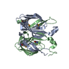

 Mass: 18.015 Da / Num. of mol.: 5 / Source method: isolated from a natural source / Formula: H2O
 Sample preparation
 Processing