 Movie
Movie Controller

[English] 日本語
 Yorodumi
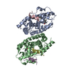
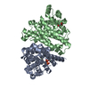
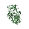
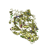
- PDB-2bc9: Crystal-structure of the N-terminal large GTPase Domain of human ... -
+ Open data

- Basic information
| Entry | Database: PDB / ID: 2bc9 | |||||||||
|---|---|---|---|---|---|---|---|---|---|---|
| Title | Crystal-structure of the N-terminal large GTPase Domain of human Guanylate Binding protein 1 (hGBP1) in complex with non-hydrolysable GTP analogue GppNHp | |||||||||
 Components Components | Interferon-induced guanylate-binding protein 1 | |||||||||
 Keywords Keywords | SIGNALING PROTEIN / Protein- guanine nucleotide complex | |||||||||
| Function / homology |  Function and homology information Function and homology informationGDP phosphatase activity / negative regulation of substrate adhesion-dependent cell spreading / non-canonical inflammasome complex assembly / protein localization to vacuole / cytolysis in another organism / positive regulation of pyroptotic inflammatory response / symbiont cell surface / vesicle membrane / negative regulation of interleukin-2 production / spectrin binding ...GDP phosphatase activity / negative regulation of substrate adhesion-dependent cell spreading / non-canonical inflammasome complex assembly / protein localization to vacuole / cytolysis in another organism / positive regulation of pyroptotic inflammatory response / symbiont cell surface / vesicle membrane / negative regulation of interleukin-2 production / spectrin binding / defense response to protozoan / cytokine binding / cellular response to interleukin-1 / negative regulation of T cell receptor signaling pathway / negative regulation of protein localization to plasma membrane / regulation of protein localization to plasma membrane / Hydrolases; Acting on acid anhydrides; In phosphorus-containing anhydrides / regulation of calcium-mediated signaling / cytoplasmic vesicle membrane / lipopolysaccharide binding / Hsp90 protein binding / negative regulation of ERK1 and ERK2 cascade / cellular response to type II interferon / cellular response to tumor necrosis factor / Interferon gamma signaling / GDP binding / actin cytoskeleton / actin binding / G protein activity / cytoplasmic vesicle / defense response to virus / Hydrolases; Acting on acid anhydrides; Acting on GTP to facilitate cellular and subcellular movement / defense response to bacterium / Golgi membrane / innate immune response / GTPase activity / GTP binding / enzyme binding / Golgi apparatus / protein homodimerization activity / extracellular region / identical protein binding / plasma membrane / cytoplasm / cytosol Similarity search - Function | |||||||||
| Biological species |  Homo sapiens (human) Homo sapiens (human) | |||||||||
| Method |  X-RAY DIFFRACTION / SYNCHROTRON / MOLECULAR REPLACEMENT / Resolution: 2.8 Å X-RAY DIFFRACTION / SYNCHROTRON / MOLECULAR REPLACEMENT / Resolution: 2.8 Å | |||||||||
 Authors Authors | Ghosh, A. / Praefcke, G.J.K. / Renault, L. / Wittinghofer, A. / Herrmann, C. | |||||||||
 Citation Citation | Journal: Nature / Year: 2006 Title: How guanylate-binding proteins achieve assembly-stimulated processive cleavage of GTP to GMP. Authors: Ghosh, A. / Praefcke, G.J. / Renault, L. / Wittinghofer, A. / Herrmann, C. #1: Journal: Embo J. / Year: 2000Title: Triphosphate structure of guanylate-binding protein 1 and implications for nucleotide binding and GTPase mechanism. Authors: Prakash, B. / Renault, L. / Praefcke, G.J. / Herrmann, C. / Wittinghofer, A. #2: Journal: Nature / Year: 2000Title: Structure of human guanylate-binding protein 1 representing a unique class of GTP-binding proteins. Authors: Prakash, B. / Praefcke, G.J. / Renault, L. / Herrmann, C. / Wittinghofer, A. | |||||||||
| History |
|
- Structure visualization
| Structure viewer | Molecule: MolmilJmol/JSmol |
|---|
- Downloads & links
-Download
| PDBx/mmCIF format | 2bc9.cif.gz | 74.9 KB | Display | PDBx/mmCIF format |
|---|---|---|---|---|
| PDB format | pdb2bc9.ent.gz | 53.9 KB | Display | PDB format |
| PDBx/mmJSON format | 2bc9.json.gz | Tree view | PDBx/mmJSON format | |
| Others |  Other downloads Other downloads |
-Validation report
| Arichive directory | https://data.pdbj.org/pub/pdb/validation_reports/bc/2bc9ftp://data.pdbj.org/pub/pdb/validation_reports/bc/2bc9 | HTTPS FTP |
|---|
-Related structure data
| Related structure data | 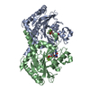 2b8wSC 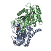 2b92C 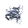 2d4hC S: Starting model for refinement C: citing same article ( |
|---|---|
| Similar structure data |


-Links
 PDBj
PDBj


- Assembly
| Deposited unit | 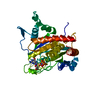
| ||||||||
|---|---|---|---|---|---|---|---|---|---|
| 1 | 
| ||||||||
| Unit cell |
| ||||||||
| Details | One of the subunit of homodimeric biological assembly is in the asymmetric unit |
-Components
| #1: Protein | Mass: 37078.449 Da / Num. of mol.: 1 / Fragment: N-terminal Large GTPase doamin Source method: isolated from a genetically manipulated source Source: (gene. exp.) Homo sapiens (human) / Gene: GBP1 / Plasmid: pQE9 / Species (production host): Escherichia coli / Production host:  |
|---|---|
| #2: Chemical | ChemComp-MG /   Mass: 24.305 Da / Num. of mol.: 1 / Source method: obtained synthetically / Formula: Mg Mass: 24.305 Da / Num. of mol.: 1 / Source method: obtained synthetically / Formula: Mg |
| #3: Chemical | ChemComp-GNP /   Mass: 522.196 Da / Num. of mol.: 1 / Source method: obtained synthetically / Formula: C10H17N6O13P3 Mass: 522.196 Da / Num. of mol.: 1 / Source method: obtained synthetically / Formula: C10H17N6O13P3Comment: GppNHp, GMPPNP, energy-carrying molecule analogue*YM |
| #4: Water | ChemComp-HOH /  Mass: 18.015 Da / Num. of mol.: 38 / Source method: isolated from a natural source / Formula: H2O Mass: 18.015 Da / Num. of mol.: 38 / Source method: isolated from a natural source / Formula: H2O |
-Experimental details
-Experiment
| Experiment | Method: X-RAY DIFFRACTION / Number of used crystals: 1 |
|---|
- Sample preparation
| Crystal | Density Matthews: 2.79 Å3/Da / Density % sol: 55.93 % |
|---|---|
| Crystal grow | Temperature: 298 K / Method: vapor diffusion, hanging drop Details: 10 % Peg 3350, 100 mM NaSCN, 40 mM CaCl2, VAPOR DIFFUSION, HANGING DROP, temperature 298K |
-Data collection
| Diffraction | Mean temperature: 100 K |
|---|---|
| Diffraction source | Source: SYNCHROTRON / Site: ESRF  / Beamline: ID14-1 / Wavelength: 0.934 Å / Beamline: ID14-1 / Wavelength: 0.934 Å |
| Detector | Type: ADSC QUANTUM 4 / Detector: CCD / Date: Nov 10, 2002 / Details: Ge (220) Crystal |
| Radiation | Monochromator: Diamond monochromator / Protocol: SINGLE WAVELENGTH / Monochromatic (M) / Laue (L): M / Scattering type: x-ray |
| Radiation wavelength | Wavelength: 0.934 Å / Relative weight: 1 |
| Reflection | Resolution: 2.8→50 Å / Num. all: 10831 / Num. obs: 10593 / % possible obs: 99.3 % / Observed criterion σ(F): 0 / Observed criterion σ(I): -3 |
| Reflection shell | Resolution: 2.8→2.9 Å / % possible all: 98.7 |
- Processing
| Software |
| ||||||||||||||||||||
|---|---|---|---|---|---|---|---|---|---|---|---|---|---|---|---|---|---|---|---|---|---|
| Refinement | Method to determine structure: MOLECULAR REPLACEMENT Starting model: PDB Entry: 2B8W Resolution: 2.8→20 Å / σ(F): 0 / Stereochemistry target values: Engh & Huber
| ||||||||||||||||||||
| Refinement step | Cycle: LAST / Resolution: 2.8→20 Å
| ||||||||||||||||||||
| Refine LS restraints |
| ||||||||||||||||||||
| LS refinement shell | Resolution: 2.8→2.9 Å /
|
