 Movie
Movie Controller

[English] 日本語
 Yorodumi
- PDB-6wkh: Crystal structure of pentalenene synthase mutant F76W complexed w... -
+ Open data

- Basic information
| Entry | Database: PDB / ID: 6wkh | ||||||
|---|---|---|---|---|---|---|---|
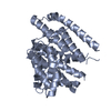
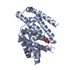
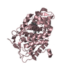
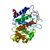
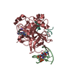
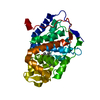
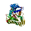
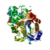
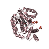
| Title | Crystal structure of pentalenene synthase mutant F76W complexed with 12,13-difluorofarnesyl diphosphate | ||||||
 Components Components | Pentalenene synthase | ||||||
 Keywords Keywords | METAL BINDING PROTEIN / LYASE/INHIBITOR / Terpene synthase / terpene synthase fold / sesquiterpene / fluoro analog / metal binding / LYASE-INHIBITOR complex | ||||||
| Function / homology |  Function and homology information Function and homology informationpentalenene synthase / pentalenene synthase activity / antibiotic biosynthetic process / metal ion binding Similarity search - Function | ||||||
| Biological species |  Streptomyces sp. (bacteria) Streptomyces sp. (bacteria) | ||||||
| Method |  X-RAY DIFFRACTION / SYNCHROTRON / MOLECULAR REPLACEMENT / Resolution: 2.55 Å X-RAY DIFFRACTION / SYNCHROTRON / MOLECULAR REPLACEMENT / Resolution: 2.55 Å | ||||||
 Authors Authors | Prem Kumar, R. / Matos, J.O. / Oprian, D.D. | ||||||
| Funding support |  United States, 1items United States, 1items
| ||||||
 Citation Citation | Journal: Biochemistry / Year: 2020 Title: Mechanism Underlying Anti-Markovnikov Addition in the Reaction of Pentalenene Synthase. Authors: Matos, J.O. / Kumar, R.P. / Ma, A.C. / Patterson, M. / Krauss, I.J. / Oprian, D.D. | ||||||
| History |
|
- Structure visualization
| Structure viewer | Molecule: MolmilJmol/JSmol |
|---|
- Downloads & links
-Download
| PDBx/mmCIF format | 6wkh.cif.gz | 125.8 KB | Display | PDBx/mmCIF format |
|---|---|---|---|---|
| PDB format | pdb6wkh.ent.gz | 96.5 KB | Display | PDB format |
| PDBx/mmJSON format | 6wkh.json.gz | Tree view | PDBx/mmJSON format | |
| Others |  Other downloads Other downloads |
-Validation report
| Arichive directory | https://data.pdbj.org/pub/pdb/validation_reports/wk/6wkhftp://data.pdbj.org/pub/pdb/validation_reports/wk/6wkh | HTTPS FTP |
|---|
-Related structure data
| Related structure data | 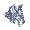 6wkcC  6wkdC  6wkeC 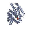 6wkfC 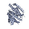 6wkgC  6wkiC  6wkjC  1ps1S S: Starting model for refinement C: citing same article ( |
|---|---|
| Similar structure data |

-Links
 PDBj
PDBj
- Assembly
| Deposited unit | 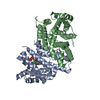
| ||||||||
|---|---|---|---|---|---|---|---|---|---|
| 1 | 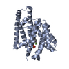
| ||||||||
| 2 | 
| ||||||||
| Unit cell |
|
-Components
| #1: Protein | Mass: 38089.445 Da / Num. of mol.: 2 / Mutation: F76W Source method: isolated from a genetically manipulated source Source: (gene. exp.) Streptomyces sp. (bacteria) / Gene: penA / Plasmid: pET28a / Production host: #2: Chemical | ChemComp-FDF / ( |   Mass: 418.307 Da / Num. of mol.: 1 / Source method: obtained synthetically / Formula: C15H26F2O7P2 / Feature type: SUBJECT OF INVESTIGATION Mass: 418.307 Da / Num. of mol.: 1 / Source method: obtained synthetically / Formula: C15H26F2O7P2 / Feature type: SUBJECT OF INVESTIGATION#3: Water | ChemComp-HOH / |  Mass: 18.015 Da / Num. of mol.: 101 / Source method: isolated from a natural source / Formula: H2O Mass: 18.015 Da / Num. of mol.: 101 / Source method: isolated from a natural source / Formula: H2OHas ligand of interest | Y | |
|---|
-Experimental details
-Experiment
| Experiment | Method: X-RAY DIFFRACTION / Number of used crystals: 1 |
|---|
- Sample preparation
| Crystal | Density Matthews: 3.69 Å3/Da / Density % sol: 66.69 % |
|---|---|
| Crystal grow | Temperature: 296 K / Method: vapor diffusion, sitting drop Details: 0.8-1.2 M sodium tartrate, 100 mM Tris, and 5 mM DTT PH range: 7.5 - 9.0 |
-Data collection
| Diffraction | Mean temperature: 100 K / Serial crystal experiment: N |
|---|---|
| Diffraction source | Source: SYNCHROTRON / Site: ALS / Beamline: 8.2.2 / Wavelength: 0.97 Å |
| Detector | Type: ADSC QUANTUM 315 / Detector: CCD / Date: Aug 16, 2019 / Details: Mirrors |
| Radiation | Monochromator: Double-crystal, Si(111) / Protocol: SINGLE WAVELENGTH / Monochromatic (M) / Laue (L): M / Scattering type: x-ray |
| Radiation wavelength | Wavelength: 0.97 Å / Relative weight: 1 |
| Reflection | Resolution: 2.55→20 Å / Num. obs: 36655 / % possible obs: 99.8 % / Redundancy: 19.8 % / CC1/2: 0.99 / Rmerge(I) obs: 0.137 / Net I/σ(I): 15.9 |
| Reflection shell | Resolution: 2.55→2.69 Å / Redundancy: 20.2 % / Rmerge(I) obs: 1.67 / Mean I/σ(I) obs: 2.1 / Num. unique obs: 5340 / % possible all: 100 |
- Processing
| Software |
| ||||||||||||||||||||||||||||||||||||||||||||||||||||||||||||||||||||||
|---|---|---|---|---|---|---|---|---|---|---|---|---|---|---|---|---|---|---|---|---|---|---|---|---|---|---|---|---|---|---|---|---|---|---|---|---|---|---|---|---|---|---|---|---|---|---|---|---|---|---|---|---|---|---|---|---|---|---|---|---|---|---|---|---|---|---|---|---|---|---|---|
| Refinement | Method to determine structure: MOLECULAR REPLACEMENT Starting model: 1PS1 Resolution: 2.55→19.886 Å / SU ML: 0.35 / Cross valid method: THROUGHOUT / σ(F): 1.34 / Phase error: 28.23 / Stereochemistry target values: ML
| ||||||||||||||||||||||||||||||||||||||||||||||||||||||||||||||||||||||
| Solvent computation | Shrinkage radii: 0.9 Å / VDW probe radii: 1.11 Å / Solvent model: FLAT BULK SOLVENT MODEL | ||||||||||||||||||||||||||||||||||||||||||||||||||||||||||||||||||||||
| Displacement parameters | Biso max: 152.81 Å2 / Biso mean: 72.7236 Å2 / Biso min: 34.97 Å2 | ||||||||||||||||||||||||||||||||||||||||||||||||||||||||||||||||||||||
| Refinement step | Cycle: final / Resolution: 2.55→19.886 Å
| ||||||||||||||||||||||||||||||||||||||||||||||||||||||||||||||||||||||
| Refine LS restraints |
| ||||||||||||||||||||||||||||||||||||||||||||||||||||||||||||||||||||||
| LS refinement shell | Refine-ID: X-RAY DIFFRACTION / Rfactor Rfree error: 0 / % reflection obs: 100 %
|
