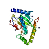
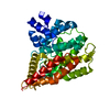
E2 ubiquitin-conjugating enzyme / ubiquitin conjugating enzyme activity / ribosomal large subunit export from nucleus / protein monoubiquitination / protein K48-linked ubiquitination / Synthesis of active ubiquitin: roles of E1 and E2 enzymes / modification-dependent protein catabolic process / protein tag activity / protein polyubiquitination / ubiquitin-protein transferase activity ...E2 ubiquitin-conjugating enzyme / ubiquitin conjugating enzyme activity / ribosomal large subunit export from nucleus / protein monoubiquitination / protein K48-linked ubiquitination / Synthesis of active ubiquitin: roles of E1 and E2 enzymes / modification-dependent protein catabolic process / protein tag activity / protein polyubiquitination / ubiquitin-protein transferase activity / Antigen processing: Ubiquitination & Proteasome degradation / ribosome biogenesis / ribosomal large subunit assembly / cytosolic large ribosomal subunit / ubiquitin-dependent protein catabolic process / cytoplasmic translation / structural constituent of ribosome / protein ubiquitination / ubiquitin protein ligase binding / ATP binding / nucleus / cytoplasm / cytosol Similarity search - Function
: / Ubiquitin Conjugating Enzyme / Ubiquitin Conjugating Enzyme / Ubiquitin-conjugating enzyme, active site / Ubiquitin-conjugating (UBC) active site signature. / Ubiquitin-conjugating enzyme E2 / Ubiquitin-conjugating enzyme / Ubiquitin-conjugating (UBC) core domain profile. / Ubiquitin-conjugating enzyme E2, catalytic domain homologues / Ubiquitin-conjugating enzyme/RWD-like ...: / Ubiquitin Conjugating Enzyme / Ubiquitin Conjugating Enzyme / Ubiquitin-conjugating enzyme, active site / Ubiquitin-conjugating (UBC) active site signature. / Ubiquitin-conjugating enzyme E2 / Ubiquitin-conjugating enzyme / Ubiquitin-conjugating (UBC) core domain profile. / Ubiquitin-conjugating enzyme E2, catalytic domain homologues / Ubiquitin-conjugating enzyme/RWD-like / Ribosomal L40e family / Ribosomal_L40e / Ribosomal protein L40e / Ribosomal protein L40e superfamily / : / Ubiquitin domain signature. / Ubiquitin conserved site / Ubiquitin domain / Ubiquitin family / Ubiquitin homologues / Ubiquitin domain profile. / Ubiquitin-like domain / Ubiquitin-like domain superfamily / Roll / Alpha Beta Similarity search - Domain/homology
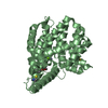
PHOSPHATE ION / Chem-U94 / Ubiquitin-ribosomal protein eL40A fusion protein / Ubiquitin-conjugating enzyme E2 R2 Similarity search - Component
Biological species
Homo sapiens (human) Schizosaccharomyces pombe (fission yeast)
In the structure databanks used in Yorodumi, some data are registered as the other names, "COVID-19 virus" and "2019-nCoV". Here are the details of the virus and the list of structure data.
Jan 31, 2019. EMDB accession codes are about to change! (news from PDBe EMDB page)
EMDB accession codes are about to change! (news from PDBe EMDB page)
The allocation of 4 digits for EMDB accession codes will soon come to an end. Whilst these codes will remain in use, new EMDB accession codes will include an additional digit and will expand incrementally as the available range of codes is exhausted. The current 4-digit format prefixed with “EMD-” (i.e. EMD-XXXX) will advance to a 5-digit format (i.e. EMD-XXXXX), and so on. It is currently estimated that the 4-digit codes will be depleted around Spring 2019, at which point the 5-digit format will come into force.
The EM Navigator/Yorodumi systems omit the EMD- prefix.
Related info.:Q: What is EMD? / ID/Accession-code notation in Yorodumi/EM Navigator
Yorodumi is a browser for structure data from EMDB, PDB, SASBDB, etc.
This page is also the successor to EM Navigator detail page, and also detail information page/front-end page for Omokage search.
The word "yorodu" (or yorozu) is an old Japanese word meaning "ten thousand". "mi" (miru) is to see.
Related info.:EMDB / PDB / SASBDB / Comparison of 3 databanks / Yorodumi Search / Aug 31, 2016. New EM Navigator & Yorodumi / Yorodumi Papers / Jmol/JSmol / Function and homology information / Changes in new EM Navigator and Yorodumi
 Movie
Movie Controller


 Open data
 Basic information
 Components
 Keywords
 Function and homology information
 Homo sapiens (human)
 X-RAY DIFFRACTION /
  Authors
 United States, 1items
  Citation
 Structure visualization
 Downloads & links
 Other downloads
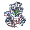


 PDBj
PDBj


 Assembly


 Mass: 442.290 Da / Num. of mol.: 1 / Source method: obtained synthetically / Formula: C20H21Cl2NO6
 Mass: 62.068 Da / Num. of mol.: 3 / Source method: obtained synthetically / Formula: C2H6O2
 Mass: 94.971 Da / Num. of mol.: 1 / Source method: obtained synthetically / Formula: PO4
 Sample preparation
 Processing