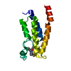
 Movie
Movie Controller


+ Open data

- Basic information
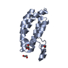
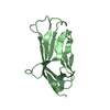
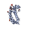
| Entry | Database: PDB / ID: 6nyc | |||||||||
|---|---|---|---|---|---|---|---|---|---|---|
| Title | Munc13-1 C2B-domain, calcium free | |||||||||
 Components Components | Munc13-1 | |||||||||
 Keywords Keywords | METAL BINDING PROTEIN / PHOSPHOLIPID BINDING PROTEIN | |||||||||
| Function / homology |  Function and homology information Function and homology informationdense core granule priming / neuronal dense core vesicle exocytosis / diacylglycerol binding / regulation of calcium-dependent activation of synaptic vesicle fusion / synaptic vesicle maturation / regulation of synaptic vesicle priming / : / positive regulation of neurotransmitter secretion / positive regulation of vesicle fusion / positive regulation of dendrite extension ...dense core granule priming / neuronal dense core vesicle exocytosis / diacylglycerol binding / regulation of calcium-dependent activation of synaptic vesicle fusion / synaptic vesicle maturation / regulation of synaptic vesicle priming / : / positive regulation of neurotransmitter secretion / positive regulation of vesicle fusion / positive regulation of dendrite extension / positive regulation of synaptic plasticity / regulation of short-term neuronal synaptic plasticity / neurotransmitter secretion / innervation / regulation of amyloid precursor protein catabolic process / syntaxin binding / syntaxin-1 binding / presynaptic active zone / Golgi-associated vesicle / spectrin binding / SNARE complex assembly / positive regulation of glutamate receptor signaling pathway / synaptic vesicle priming / neuromuscular junction development / amyloid-beta metabolic process / synaptic vesicle exocytosis / excitatory synapse / calyx of Held / SNARE binding / synaptic membrane / neuromuscular junction / synaptic transmission, glutamatergic / phospholipid binding / long-term synaptic potentiation / terminal bouton / synaptic vesicle membrane / presynapse / presynaptic membrane / cell differentiation / calmodulin binding / protein domain specific binding / axon / calcium ion binding / synapse / protein-containing complex binding / glutamatergic synapse / protein-containing complex / zinc ion binding / identical protein binding / plasma membrane Similarity search - Function | |||||||||
| Biological species |  | |||||||||
| Method |  X-RAY DIFFRACTION / SYNCHROTRON / MOLECULAR REPLACEMENT / Resolution: 1.893 Å X-RAY DIFFRACTION / SYNCHROTRON / MOLECULAR REPLACEMENT / Resolution: 1.893 Å | |||||||||
 Authors Authors | Tomchick, D.R. / Rizo, J. / Machius, M. / Lu, J. | |||||||||
| Funding support |  United States, 2items United States, 2items
| |||||||||
 Citation Citation | Journal: Nat. Struct. Mol. Biol. / Year: 2010 Title: Munc13 C2B domain is an activity-dependent Ca2+ regulator of synaptic exocytosis. Authors: Shin, O.H. / Lu, J. / Rhee, J.S. / Tomchick, D.R. / Pang, Z.P. / Wojcik, S.M. / Camacho-Perez, M. / Brose, N. / Machius, M. / Rizo, J. / Rosenmund, C. / Sudhof, T.C. #1: Journal: Cell / Year: 2004 Title: Calmodulin and Munc13 form a Ca2+ sensor/effector complex that controls short-term synaptic plasticity. Authors: Junge, H.J. / Rhee, J.S. / Jahn, O. / Varoqueaux, F. / Speiss, J. / Waxham, M.N. / Rosenmund, C. / Brose, N. #2: Journal: Nature / Year: 1999 Title: Munc-13 is essential for fusion competence of glutamatergic synaptic vesicles. Authors: Augustin, I. / Rosenmund, C. / Sudhof, T.C. / Brose, N. #3: Journal: J. Biol. Chem. / Year: 1995 Title: Mammalian homologues of C. elegans unc-13 gene define novel family of C2-domain proteins. Authors: Brose, N. / Hofmann, K. / Hata, Y. / Sudhof, T.C. | |||||||||
| History |
|
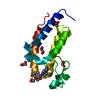
- Structure visualization
| Structure viewer | Molecule: MolmilJmol/JSmol |
|---|
- Downloads & links
-Download
| PDBx/mmCIF format | 6nyc.cif.gz | 85.6 KB | Display | PDBx/mmCIF format |
|---|---|---|---|---|
| PDB format | pdb6nyc.ent.gz | 64.2 KB | Display | PDB format |
| PDBx/mmJSON format | 6nyc.json.gz | Tree view | PDBx/mmJSON format | |
| Others |  Other downloads Other downloads |
-Validation report
| Arichive directory | https://data.pdbj.org/pub/pdb/validation_reports/ny/6nycftp://data.pdbj.org/pub/pdb/validation_reports/ny/6nyc | HTTPS FTP |
|---|
-Related structure data
| Related structure data |  6nytC  1rsyS S: Starting model for refinement C: citing same article ( |
|---|---|
| Similar structure data |


-Links
 PDBj
PDBj


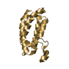
- Assembly
| Deposited unit | 
| ||||||||
|---|---|---|---|---|---|---|---|---|---|
| 1 |
| ||||||||
| Unit cell |
|
-Components
| #1: Protein | Mass: 16937.131 Da / Num. of mol.: 1 / Fragment: C2B domain, residues 675-820 / Mutation: L756W Source method: isolated from a genetically manipulated source Source: (gene. exp.)  | ||||
|---|---|---|---|---|---|
| #2: Chemical |   Mass: 35.453 Da / Num. of mol.: 2 / Source method: obtained synthetically / Formula: Cl Mass: 35.453 Da / Num. of mol.: 2 / Source method: obtained synthetically / Formula: Cl#3: Chemical | ChemComp-B3P / |   Mass: 282.334 Da / Num. of mol.: 1 / Source method: obtained synthetically / Formula: C11H26N2O6 / Comment: pH buffer*YM Mass: 282.334 Da / Num. of mol.: 1 / Source method: obtained synthetically / Formula: C11H26N2O6 / Comment: pH buffer*YM#4: Water | ChemComp-HOH / |  Mass: 18.015 Da / Num. of mol.: 20 / Source method: isolated from a natural source / Formula: H2O Mass: 18.015 Da / Num. of mol.: 20 / Source method: isolated from a natural source / Formula: H2O |
-Experimental details
-Experiment
| Experiment | Method: X-RAY DIFFRACTION / Number of used crystals: 1 |
|---|
- Sample preparation
| Crystal | Density Matthews: 2.16 Å3/Da / Density % sol: 43.11 % / Mosaicity: 0.635 ° |
|---|---|
| Crystal grow | Temperature: 298 K / Method: vapor diffusion, hanging drop / pH: 6.8 Details: 30% PEG-MME 2000, 0.1 M bis-tris propane pH 6.8, 0.1 M NaCl, 0.5 mM TCEP |
-Data collection
| Diffraction | Mean temperature: 100 K / Serial crystal experiment: N | |||||||||||||||||||||||||||||||||||||||||||||||||||||||||||||||||||||||||||||||||||||||||||||||||||||||||||||||||||||||||||||||||||||||||||||||||||
|---|---|---|---|---|---|---|---|---|---|---|---|---|---|---|---|---|---|---|---|---|---|---|---|---|---|---|---|---|---|---|---|---|---|---|---|---|---|---|---|---|---|---|---|---|---|---|---|---|---|---|---|---|---|---|---|---|---|---|---|---|---|---|---|---|---|---|---|---|---|---|---|---|---|---|---|---|---|---|---|---|---|---|---|---|---|---|---|---|---|---|---|---|---|---|---|---|---|---|---|---|---|---|---|---|---|---|---|---|---|---|---|---|---|---|---|---|---|---|---|---|---|---|---|---|---|---|---|---|---|---|---|---|---|---|---|---|---|---|---|---|---|---|---|---|---|---|---|---|
| Diffraction source | Source: SYNCHROTRON / Site: APS / Beamline: 19-BM / Wavelength: 0.98066 Å | |||||||||||||||||||||||||||||||||||||||||||||||||||||||||||||||||||||||||||||||||||||||||||||||||||||||||||||||||||||||||||||||||||||||||||||||||||
| Detector | Type: SBC-3 / Detector: CCD / Date: Nov 23, 2003 / Details: monochromator | |||||||||||||||||||||||||||||||||||||||||||||||||||||||||||||||||||||||||||||||||||||||||||||||||||||||||||||||||||||||||||||||||||||||||||||||||||
| Radiation | Monochromator: SAGITALLY FOCUSED Si(111) / Protocol: SINGLE WAVELENGTH / Monochromatic (M) / Laue (L): M / Scattering type: x-ray | |||||||||||||||||||||||||||||||||||||||||||||||||||||||||||||||||||||||||||||||||||||||||||||||||||||||||||||||||||||||||||||||||||||||||||||||||||
| Radiation wavelength | Wavelength: 0.98066 Å / Relative weight: 1 | |||||||||||||||||||||||||||||||||||||||||||||||||||||||||||||||||||||||||||||||||||||||||||||||||||||||||||||||||||||||||||||||||||||||||||||||||||
| Reflection | Resolution: 1.893→50 Å / Num. obs: 10420 / % possible obs: 86.5 % / Redundancy: 7.2 % / Rmerge(I) obs: 0.05 / Χ2: 1.07 / Net I/σ(I): 17 | |||||||||||||||||||||||||||||||||||||||||||||||||||||||||||||||||||||||||||||||||||||||||||||||||||||||||||||||||||||||||||||||||||||||||||||||||||
| Reflection shell |
|
- Processing
| Software |
| ||||||||||||||||||||||||||||||||||||||||||||||||||||||||||||||||||||||||||||||||||||||||||||||||||||
|---|---|---|---|---|---|---|---|---|---|---|---|---|---|---|---|---|---|---|---|---|---|---|---|---|---|---|---|---|---|---|---|---|---|---|---|---|---|---|---|---|---|---|---|---|---|---|---|---|---|---|---|---|---|---|---|---|---|---|---|---|---|---|---|---|---|---|---|---|---|---|---|---|---|---|---|---|---|---|---|---|---|---|---|---|---|---|---|---|---|---|---|---|---|---|---|---|---|---|---|---|---|
| Refinement | Method to determine structure: MOLECULAR REPLACEMENT Starting model: 1RSY Resolution: 1.893→26.43 Å / SU ML: 0.18 / Cross valid method: THROUGHOUT / σ(F): 1.37 / Phase error: 34.31
| ||||||||||||||||||||||||||||||||||||||||||||||||||||||||||||||||||||||||||||||||||||||||||||||||||||
| Solvent computation | Shrinkage radii: 0.9 Å / VDW probe radii: 1.11 Å | ||||||||||||||||||||||||||||||||||||||||||||||||||||||||||||||||||||||||||||||||||||||||||||||||||||
| Displacement parameters | Biso max: 147.26 Å2 / Biso mean: 52.568 Å2 / Biso min: 20.37 Å2 | ||||||||||||||||||||||||||||||||||||||||||||||||||||||||||||||||||||||||||||||||||||||||||||||||||||
| Refinement step | Cycle: final / Resolution: 1.893→26.43 Å
| ||||||||||||||||||||||||||||||||||||||||||||||||||||||||||||||||||||||||||||||||||||||||||||||||||||
| LS refinement shell | Refine-ID: X-RAY DIFFRACTION / Rfactor Rfree error: 0 / Total num. of bins used: 4
| ||||||||||||||||||||||||||||||||||||||||||||||||||||||||||||||||||||||||||||||||||||||||||||||||||||
| Refinement TLS params. | Method: refined / Refine-ID: X-RAY DIFFRACTION
| ||||||||||||||||||||||||||||||||||||||||||||||||||||||||||||||||||||||||||||||||||||||||||||||||||||
| Refinement TLS group |
|
