 Movie
Movie Controller

[English] 日本語
 Yorodumi
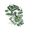
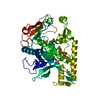
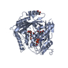
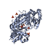
- PDB-6ni5: Pseudomonas fluorescens isocyanide hydratase at 274 K G150A mutant -
+ Open data

- Basic information
| Entry | Database: PDB / ID: 6ni5 | |||||||||
|---|---|---|---|---|---|---|---|---|---|---|
| Title | Pseudomonas fluorescens isocyanide hydratase at 274 K G150A mutant | |||||||||
 Components Components | Isonitrile hydratase InhA | |||||||||
 Keywords Keywords | LYASE / DJ-1 superfamily | |||||||||
| Function / homology |  Function and homology information Function and homology information | |||||||||
| Biological species |  Pseudomonas fluorescens (bacteria) Pseudomonas fluorescens (bacteria) | |||||||||
| Method |  X-RAY DIFFRACTION / SYNCHROTRON / MOLECULAR REPLACEMENT / Resolution: 1.3 Å X-RAY DIFFRACTION / SYNCHROTRON / MOLECULAR REPLACEMENT / Resolution: 1.3 Å | |||||||||
 Authors Authors | Wilson, M.A. / Dasgupta, M. / van den Bedem, H. | |||||||||
| Funding support |  United States, 1items United States, 1items
| |||||||||
 Citation Citation | Journal: Proc.Natl.Acad.Sci.USA / Year: 2019 Title: Mix-and-inject XFEL crystallography reveals gated conformational dynamics during enzyme catalysis. Authors: Dasgupta, M. / Budday, D. / de Oliveira, S.H.P. / Madzelan, P. / Marchany-Rivera, D. / Seravalli, J. / Hayes, B. / Sierra, R.G. / Boutet, S. / Hunter, M.S. / Alonso-Mori, R. / Batyuk, A. / ...Authors: Dasgupta, M. / Budday, D. / de Oliveira, S.H.P. / Madzelan, P. / Marchany-Rivera, D. / Seravalli, J. / Hayes, B. / Sierra, R.G. / Boutet, S. / Hunter, M.S. / Alonso-Mori, R. / Batyuk, A. / Wierman, J. / Lyubimov, A. / Brewster, A.S. / Sauter, N.K. / Applegate, G.A. / Tiwari, V.K. / Berkowitz, D.B. / Thompson, M.C. / Cohen, A.E. / Fraser, J.S. / Wall, M.E. / van den Bedem, H. / Wilson, M.A. | |||||||||
| History |
|
- Structure visualization
| Structure viewer | Molecule: MolmilJmol/JSmol |
|---|
- Downloads & links
-Download
| PDBx/mmCIF format | 6ni5.cif.gz | 358.8 KB | Display | PDBx/mmCIF format |
|---|---|---|---|---|
| PDB format | pdb6ni5.ent.gz | 306.6 KB | Display | PDB format |
| PDBx/mmJSON format | 6ni5.json.gz | Tree view | PDBx/mmJSON format | |
| Others |  Other downloads Other downloads |
-Validation report
| Arichive directory | https://data.pdbj.org/pub/pdb/validation_reports/ni/6ni5ftp://data.pdbj.org/pub/pdb/validation_reports/ni/6ni5 | HTTPS FTP |
|---|
-Related structure data
| Related structure data |  6ni4C  6ni6C  6ni7C  6ni9C  6niaC  6npqC  6undC  6unfC  3nonS S: Starting model for refinement C: citing same article ( |
|---|---|
| Similar structure data |


-Links
 PDBj
PDBj

- Assembly
| Deposited unit | 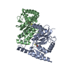
| ||||||||
|---|---|---|---|---|---|---|---|---|---|
| 1 |
| ||||||||
| Unit cell |
|
-Components
| #1: Protein | Mass: 24194.674 Da / Num. of mol.: 2 / Mutation: G150A Source method: isolated from a genetically manipulated source Source: (gene. exp.) Pseudomonas fluorescens (strain ATCC BAA-477 / NRRL B-23932 / Pf-5) (bacteria)Strain: ATCC BAA-477 / NRRL B-23932 / Pf-5 / Gene: inhA, PFL_4109 / Plasmid: pET15b / Production host: #2: Water | ChemComp-HOH / |  Mass: 18.015 Da / Num. of mol.: 323 / Source method: isolated from a natural source / Formula: H2O Mass: 18.015 Da / Num. of mol.: 323 / Source method: isolated from a natural source / Formula: H2OHas protein modification | Y | |
|---|
-Experimental details
-Experiment
| Experiment | Method: X-RAY DIFFRACTION / Number of used crystals: 1 |
|---|
- Sample preparation
| Crystal | Density Matthews: 2.18 Å3/Da / Density % sol: 43.52 % |
|---|---|
| Crystal grow | Temperature: 298 K / Method: vapor diffusion, hanging drop / pH: 8.6 Details: 25% PEG 3350, 200 mM magnesium chloride, 100mM Tris-HCl, pH 8.6, 2 mM DTT |
-Data collection
| Diffraction | Mean temperature: 274 K / Serial crystal experiment: N |
|---|---|
| Diffraction source | Source: SYNCHROTRON / Site: SSRL / Beamline: BL12-2 / Wavelength: 0.82653 Å |
| Detector | Type: DECTRIS PILATUS 6M / Detector: PIXEL / Date: Feb 25, 2016 Details: Flat Si Rh coated M0, Kirkpatrick-Baez flat bent Si M1 & M2 |
| Radiation | Monochromator: Liquid nitrogen-cooled double crystal Si(111) Protocol: SINGLE WAVELENGTH / Monochromatic (M) / Laue (L): M / Scattering type: x-ray |
| Radiation wavelength | Wavelength: 0.82653 Å / Relative weight: 1 |
| Reflection | Resolution: 1.3→38.96 Å / Num. obs: 100249 / % possible obs: 98.1 % / Redundancy: 3.3 % / CC1/2: 0.996 / Rmerge(I) obs: 0.055 / Rpim(I) all: 0.036 / Rrim(I) all: 0.066 / Net I/σ(I): 9.1 |
| Reflection shell | Resolution: 1.3→1.32 Å / Redundancy: 3 % / Rmerge(I) obs: 1.513 / Mean I/σ(I) obs: 1.2 / Num. unique obs: 4568 / CC1/2: 0.318 / Rpim(I) all: 0.846 / Rrim(I) all: 1.513 / % possible all: 90.1 |
- Processing
| Software |
| |||||||||||||||||||||||||||||||||||||||||||||||||||||||||||||||||||||||||||||||||||||||||||||||||||||||||||||||||||||||||||||||||||||||||||||||||||||||||||||||||||||||||||||||||||||||||||||||||||||||||||||||||||||||||
|---|---|---|---|---|---|---|---|---|---|---|---|---|---|---|---|---|---|---|---|---|---|---|---|---|---|---|---|---|---|---|---|---|---|---|---|---|---|---|---|---|---|---|---|---|---|---|---|---|---|---|---|---|---|---|---|---|---|---|---|---|---|---|---|---|---|---|---|---|---|---|---|---|---|---|---|---|---|---|---|---|---|---|---|---|---|---|---|---|---|---|---|---|---|---|---|---|---|---|---|---|---|---|---|---|---|---|---|---|---|---|---|---|---|---|---|---|---|---|---|---|---|---|---|---|---|---|---|---|---|---|---|---|---|---|---|---|---|---|---|---|---|---|---|---|---|---|---|---|---|---|---|---|---|---|---|---|---|---|---|---|---|---|---|---|---|---|---|---|---|---|---|---|---|---|---|---|---|---|---|---|---|---|---|---|---|---|---|---|---|---|---|---|---|---|---|---|---|---|---|---|---|---|---|---|---|---|---|---|---|---|---|---|---|---|---|---|---|---|
| Refinement | Method to determine structure: MOLECULAR REPLACEMENT Starting model: 3NON Resolution: 1.3→33.595 Å / SU ML: 0.14 / Cross valid method: FREE R-VALUE / σ(F): 1.34 / Phase error: 14.88 / Stereochemistry target values: ML
| |||||||||||||||||||||||||||||||||||||||||||||||||||||||||||||||||||||||||||||||||||||||||||||||||||||||||||||||||||||||||||||||||||||||||||||||||||||||||||||||||||||||||||||||||||||||||||||||||||||||||||||||||||||||||
| Solvent computation | Shrinkage radii: 0.9 Å / VDW probe radii: 1.11 Å / Solvent model: FLAT BULK SOLVENT MODEL | |||||||||||||||||||||||||||||||||||||||||||||||||||||||||||||||||||||||||||||||||||||||||||||||||||||||||||||||||||||||||||||||||||||||||||||||||||||||||||||||||||||||||||||||||||||||||||||||||||||||||||||||||||||||||
| Refinement step | Cycle: LAST / Resolution: 1.3→33.595 Å
| |||||||||||||||||||||||||||||||||||||||||||||||||||||||||||||||||||||||||||||||||||||||||||||||||||||||||||||||||||||||||||||||||||||||||||||||||||||||||||||||||||||||||||||||||||||||||||||||||||||||||||||||||||||||||
| Refine LS restraints |
| |||||||||||||||||||||||||||||||||||||||||||||||||||||||||||||||||||||||||||||||||||||||||||||||||||||||||||||||||||||||||||||||||||||||||||||||||||||||||||||||||||||||||||||||||||||||||||||||||||||||||||||||||||||||||
| LS refinement shell |
|
