 Movie
Movie Controller

[English] 日本語
 Yorodumi
- PDB-6ncz: Crystal structure of hybrid beta-glucuronidase/beta-galacturonida... -
+ Open data

- Basic information
| Entry | Database: PDB / ID: 6ncz | |||||||||
|---|---|---|---|---|---|---|---|---|---|---|
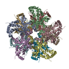
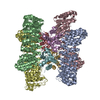
| Title | Crystal structure of hybrid beta-glucuronidase/beta-galacturonidase from Fusicatenibacter saccharivorans bound to phenyl-thio-beta-D-glucuronide | |||||||||
 Components Components | Beta-glucuronidase | |||||||||
 Keywords Keywords | HYDROLASE / glycoside hydrolase family 2 / beta-glucuronidase / beta-galacturonidase | |||||||||
| Function / homology |  Function and homology information Function and homology informationglucuronoside catabolic process / beta-glucuronidase / beta-glucuronidase activity / carbohydrate binding / carbohydrate metabolic process Similarity search - Function | |||||||||
| Biological species |  Fusicatenibacter saccharivorans (bacteria) Fusicatenibacter saccharivorans (bacteria) | |||||||||
| Method |  X-RAY DIFFRACTION / SYNCHROTRON / MOLECULAR REPLACEMENT / Resolution: 2.2 Å X-RAY DIFFRACTION / SYNCHROTRON / MOLECULAR REPLACEMENT / Resolution: 2.2 Å | |||||||||
 Authors Authors | Walton, W.G. / Pellock, S.J. / Redinbo, M.R. | |||||||||
| Funding support |  United States, 1items United States, 1items
| |||||||||
 Citation Citation | Journal: Biochemistry / Year: 2019 Title: Selecting a Single Stereocenter: The Molecular Nuances That Differentiate beta-Hexuronidases in the Human Gut Microbiome. Authors: Pellock, S.J. / Walton, W.G. / Redinbo, M.R. | |||||||||
| History |
|
- Structure visualization
| Structure viewer | Molecule: MolmilJmol/JSmol |
|---|
- Downloads & links
-Download
| PDBx/mmCIF format | 6ncz.cif.gz | 744.1 KB | Display | PDBx/mmCIF format |
|---|---|---|---|---|
| PDB format | pdb6ncz.ent.gz | 613.4 KB | Display | PDB format |
| PDBx/mmJSON format | 6ncz.json.gz | Tree view | PDBx/mmJSON format | |
| Others |  Other downloads Other downloads |
-Validation report
| Arichive directory | https://data.pdbj.org/pub/pdb/validation_reports/nc/6nczftp://data.pdbj.org/pub/pdb/validation_reports/nc/6ncz | HTTPS FTP |
|---|
-Related structure data
| Related structure data |  6ncwC  6ncxC 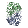 6ncyC 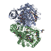 3lpfS S: Starting model for refinement C: citing same article ( |
|---|---|
| Similar structure data |


-Links
 PDBj
PDBj

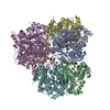
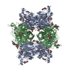
- Assembly
| Deposited unit | 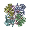
| ||||||||
|---|---|---|---|---|---|---|---|---|---|
| 1 |
| ||||||||
| 2 | 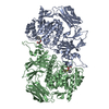
| ||||||||
| 3 | 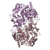
| ||||||||
| 4 | 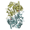
| ||||||||
| Unit cell |
|
-Components
| #1: Protein | Mass: 71263.914 Da / Num. of mol.: 6 Source method: isolated from a genetically manipulated source Source: (gene. exp.) Fusicatenibacter saccharivorans (bacteria)Gene: uidA_2, ERS852406_01826 / Production host: #2: Sugar | ChemComp-FYJ / 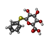  Type: D-saccharide / Mass: 286.301 Da / Num. of mol.: 6 / Source method: obtained synthetically / Formula: C12H14O6S Type: D-saccharide / Mass: 286.301 Da / Num. of mol.: 6 / Source method: obtained synthetically / Formula: C12H14O6S#3: Chemical | ChemComp-GOL /   Mass: 92.094 Da / Num. of mol.: 6 / Source method: obtained synthetically / Formula: C3H8O3 Mass: 92.094 Da / Num. of mol.: 6 / Source method: obtained synthetically / Formula: C3H8O3#4: Water | ChemComp-HOH / |  Mass: 18.015 Da / Num. of mol.: 1768 / Source method: isolated from a natural source / Formula: H2O Mass: 18.015 Da / Num. of mol.: 1768 / Source method: isolated from a natural source / Formula: H2O |
|---|
-Experimental details
-Experiment
| Experiment | Method: X-RAY DIFFRACTION / Number of used crystals: 1 |
|---|
- Sample preparation
| Crystal | Density Matthews: 2.63 Å3/Da / Density % sol: 53.27 % |
|---|---|
| Crystal grow | Temperature: 293 K / Method: vapor diffusion, sitting drop Details: 0.2 M sodium tartrate dibasic, 20% (w/v) PEG 3350 with 1 mM phenyl-thio-beta-D-glucuronide |
-Data collection
| Diffraction | Mean temperature: 100 K / Serial crystal experiment: N |
|---|---|
| Diffraction source | Source: SYNCHROTRON / Site: APS / Beamline: 23-ID-D / Wavelength: 1.033 Å |
| Detector | Type: DECTRIS PILATUS3 6M / Detector: PIXEL / Date: Jun 10, 2018 |
| Radiation | Protocol: SINGLE WAVELENGTH / Monochromatic (M) / Laue (L): M / Scattering type: x-ray |
| Radiation wavelength | Wavelength: 1.033 Å / Relative weight: 1 |
| Reflection | Resolution: 2.2→29.7 Å / Num. obs: 215731 / % possible obs: 97.4 % / Redundancy: 3.6 % / CC1/2: 0.995 / Rmerge(I) obs: 0.116 / Rpim(I) all: 0.071 / Net I/σ(I): 8.3 |
| Reflection shell | Resolution: 2.2→2.28 Å / Redundancy: 3.6 % / Rmerge(I) obs: 1.093 / Mean I/σ(I) obs: 1.1 / Num. unique obs: 21435 / CC1/2: 0.498 / Rpim(I) all: 0.664 / % possible all: 96.8 |
- Processing
| Software |
| |||||||||||||||||||||||||||||||||||||||||||||||||||||||||||||||||||||||||||||||||||||||||||||||||||||||||
|---|---|---|---|---|---|---|---|---|---|---|---|---|---|---|---|---|---|---|---|---|---|---|---|---|---|---|---|---|---|---|---|---|---|---|---|---|---|---|---|---|---|---|---|---|---|---|---|---|---|---|---|---|---|---|---|---|---|---|---|---|---|---|---|---|---|---|---|---|---|---|---|---|---|---|---|---|---|---|---|---|---|---|---|---|---|---|---|---|---|---|---|---|---|---|---|---|---|---|---|---|---|---|---|---|---|---|
| Refinement | Method to determine structure: MOLECULAR REPLACEMENT Starting model: 3LPF Resolution: 2.2→29.67 Å / SU ML: 0.3 / Cross valid method: FREE R-VALUE / σ(F): 1.96 / Phase error: 25.3 / Stereochemistry target values: ML
| |||||||||||||||||||||||||||||||||||||||||||||||||||||||||||||||||||||||||||||||||||||||||||||||||||||||||
| Solvent computation | Shrinkage radii: 0.9 Å / VDW probe radii: 1.11 Å / Solvent model: FLAT BULK SOLVENT MODEL | |||||||||||||||||||||||||||||||||||||||||||||||||||||||||||||||||||||||||||||||||||||||||||||||||||||||||
| Refinement step | Cycle: LAST / Resolution: 2.2→29.67 Å
| |||||||||||||||||||||||||||||||||||||||||||||||||||||||||||||||||||||||||||||||||||||||||||||||||||||||||
| Refine LS restraints |
| |||||||||||||||||||||||||||||||||||||||||||||||||||||||||||||||||||||||||||||||||||||||||||||||||||||||||
| LS refinement shell |
|
