 Movie
Movie Controller

[English] 日本語
 Yorodumi
- PDB-1v3c: Structure of the hemagglutinin-neuraminidase from human parainflu... -
+ Open data

- Basic information
| Entry | Database: PDB / ID: 1v3c | |||||||||
|---|---|---|---|---|---|---|---|---|---|---|
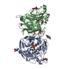
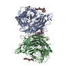
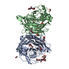
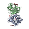
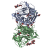
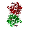
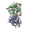
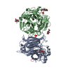
| Title | Structure of the hemagglutinin-neuraminidase from human parainfluenza virus type III: complex with NEU5AC | |||||||||
 Components Components | hemagglutinin-neuraminidase glycoprotein | |||||||||
 Keywords Keywords | HYDROLASE / PIV3 HN / NATIVE+NEU5AC / HEXAGONAL | |||||||||
| Function / homology |  Function and homology information Function and homology informationexo-alpha-sialidase activity / host cell surface receptor binding / viral envelope / symbiont entry into host cell / virion attachment to host cell / host cell plasma membrane / virion membrane / metal ion binding Similarity search - Function | |||||||||
| Biological species |  Human parainfluenza virus 3 Human parainfluenza virus 3 | |||||||||
| Method |  X-RAY DIFFRACTION / SYNCHROTRON / MOLECULAR REPLACEMENT / Resolution: 2.3 Å X-RAY DIFFRACTION / SYNCHROTRON / MOLECULAR REPLACEMENT / Resolution: 2.3 Å | |||||||||
 Authors Authors | Lawrence, M.C. / Borg, N.A. / Streltsov, V.A. / Pilling, P.A. / Epa, V.C. / Varghese, J.N. / McKimm-Breschkin, J.L. / Colman, P.M. | |||||||||
 Citation Citation | Journal: J.Mol.Biol. / Year: 2004 Title: Structure of the Haemagglutinin-neuraminidase from Human Parainfluenza Virus Type III Authors: Lawrence, M.C. / Borg, N.A. / Streltsov, V.A. / Pilling, P.A. / Epa, V.C. / Varghese, J.N. / McKimm-Breschkin, J.L. / Colman, P.M. | |||||||||
| History |
|
- Structure visualization
| Structure viewer | Molecule: MolmilJmol/JSmol |
|---|
- Downloads & links
-Download
| PDBx/mmCIF format | 1v3c.cif.gz | 207.3 KB | Display | PDBx/mmCIF format |
|---|---|---|---|---|
| PDB format | pdb1v3c.ent.gz | 162.8 KB | Display | PDB format |
| PDBx/mmJSON format | 1v3c.json.gz | Tree view | PDBx/mmJSON format | |
| Others |  Other downloads Other downloads |
-Validation report
| Arichive directory | https://data.pdbj.org/pub/pdb/validation_reports/v3/1v3cftp://data.pdbj.org/pub/pdb/validation_reports/v3/1v3c | HTTPS FTP |
|---|
-Related structure data
| Related structure data | 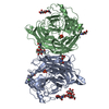 1v2iC  1v3bSC  1v3dC 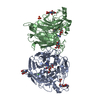 1v3eC C: citing same article ( S: Starting model for refinement |
|---|---|
| Similar structure data |


-Links
 PDBj
PDBj

- Assembly
| Deposited unit | 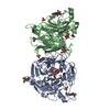
| ||||||||||||
|---|---|---|---|---|---|---|---|---|---|---|---|---|---|
| 1 |
| ||||||||||||
| 2 | 
| ||||||||||||
| Unit cell |
| ||||||||||||
| Components on special symmetry positions |
|
-Components
-Protein , 1 types, 2 molecules AB
| #1: Protein | Mass: 48107.715 Da / Num. of mol.: 2 / Fragment: RESIDUES 142-572 Source method: isolated from a genetically manipulated source Source: (gene. exp.) Human parainfluenza virus 3 / Genus: Respirovirus / Cell line (production host): HIGH FIVE / Production host:  Trichoplusia ni (cabbage looper) Trichoplusia ni (cabbage looper)References: GenBank: 37958139, UniProt: Q6WJ03*PLUS, exo-alpha-sialidase |
|---|
-Sugars , 3 types, 7 molecules 
| #2: Polysaccharide | Source method: isolated from a genetically manipulated source #3: Sugar |  Type: D-saccharide, beta linking / Mass: 221.208 Da / Num. of mol.: 3 Type: D-saccharide, beta linking / Mass: 221.208 Da / Num. of mol.: 3Source method: isolated from a genetically manipulated source Formula: C8H15NO6 #4: Sugar |  Type: D-saccharide, beta linking / Mass: 309.270 Da / Num. of mol.: 2 Type: D-saccharide, beta linking / Mass: 309.270 Da / Num. of mol.: 2Source method: isolated from a genetically manipulated source Formula: C11H19NO9 |
|---|
-Non-polymers , 3 types, 840 molecules 


| #5: Chemical |  Mass: 40.078 Da / Num. of mol.: 2 / Source method: obtained synthetically / Formula: Ca Mass: 40.078 Da / Num. of mol.: 2 / Source method: obtained synthetically / Formula: Ca#6: Chemical | ChemComp-SO4 /  Mass: 96.063 Da / Num. of mol.: 4 / Source method: obtained synthetically / Formula: SO4 Mass: 96.063 Da / Num. of mol.: 4 / Source method: obtained synthetically / Formula: SO4#7: Water | ChemComp-HOH / | Mass: 18.015 Da / Num. of mol.: 834 / Source method: isolated from a natural source / Formula: H2O |
|---|
-Details
| Has protein modification | Y |
|---|
-Experimental details
-Experiment
| Experiment | Method: X-RAY DIFFRACTION / Number of used crystals: 1 |
|---|
- Sample preparation
| Crystal | Density Matthews: 3.49 Å3/Da / Density % sol: 64.46 % | |||||||||||||||||||||||||||||||||||
|---|---|---|---|---|---|---|---|---|---|---|---|---|---|---|---|---|---|---|---|---|---|---|---|---|---|---|---|---|---|---|---|---|---|---|---|---|
| Crystal grow | Temperature: 298 K / Method: vapor diffusion, hanging drop / pH: 7.5 Details: AMMONIUM AND LITHIUM PHOSPHATE, HEPES, pH 7.50, VAPOR DIFFUSION, HANGING DROP, temperature 298K | |||||||||||||||||||||||||||||||||||
| Crystal grow | *PLUS pH: 7.5 / Method: vapor diffusion | |||||||||||||||||||||||||||||||||||
| Components of the solutions | *PLUS
|
-Data collection
| Diffraction | Mean temperature: 113 K |
|---|---|
| Diffraction source | Source: SYNCHROTRON / Site: APS  / Beamline: 14-ID-B / Wavelength: 1 / Beamline: 14-ID-B / Wavelength: 1 |
| Detector | Type: MARRESEARCH / Detector: CCD |
| Radiation | Monochromator: DIAMOND 111 / Protocol: SINGLE WAVELENGTH / Monochromatic (M) / Laue (L): M / Scattering type: x-ray |
| Radiation wavelength | Wavelength: 1 Å / Relative weight: 1 |
| Reflection | Resolution: 2.3→8.5 Å / Num. obs: 61373 / % possible obs: 99.9 % / Observed criterion σ(I): 0 / Redundancy: 8.8 % / Biso Wilson estimate: 24.8 Å2 / Net I/σ(I): 12.28 |
| Reflection shell | Resolution: 2.3→2.34 Å / Redundancy: 8.1 % / Mean I/σ(I) obs: 1.9 / % possible all: 100 |
| Reflection | *PLUS Highest resolution: 2.3 Å / Redundancy: 8.8 % / Num. measured all: 539280 |
| Reflection shell | *PLUS % possible obs: 100 % / Mean I/σ(I) obs: 3.6 |
- Processing
| Software |
| ||||||||||||||||||||||||||||||||||||||||||||||||||||||||||||
|---|---|---|---|---|---|---|---|---|---|---|---|---|---|---|---|---|---|---|---|---|---|---|---|---|---|---|---|---|---|---|---|---|---|---|---|---|---|---|---|---|---|---|---|---|---|---|---|---|---|---|---|---|---|---|---|---|---|---|---|---|---|
| Refinement | Method to determine structure: MOLECULAR REPLACEMENT Starting model: PIV3 HN MONOMER FROM HEXAGONAL STRUCTURE ENTRY 1V3B Resolution: 2.3→7.5 Å / Rfactor Rfree error: 0.004 / Data cutoff high absF: 169994.91 / Data cutoff low absF: 0 / Isotropic thermal model: RESTRAINED / Cross valid method: THROUGHOUT / σ(F): 0
| ||||||||||||||||||||||||||||||||||||||||||||||||||||||||||||
| Solvent computation | Solvent model: FLAT MODEL / Bsol: 56.3363 Å2 / ksol: 0.352789 e/Å3 | ||||||||||||||||||||||||||||||||||||||||||||||||||||||||||||
| Displacement parameters | Biso mean: 31.9 Å2
| ||||||||||||||||||||||||||||||||||||||||||||||||||||||||||||
| Refine analyze |
| ||||||||||||||||||||||||||||||||||||||||||||||||||||||||||||
| Refinement step | Cycle: LAST / Resolution: 2.3→7.5 Å
| ||||||||||||||||||||||||||||||||||||||||||||||||||||||||||||
| Refine LS restraints |
| ||||||||||||||||||||||||||||||||||||||||||||||||||||||||||||
| LS refinement shell | Resolution: 2.3→2.44 Å / Rfactor Rfree error: 0.013 / Total num. of bins used: 6
| ||||||||||||||||||||||||||||||||||||||||||||||||||||||||||||
| Xplor file |
| ||||||||||||||||||||||||||||||||||||||||||||||||||||||||||||
| Refinement | *PLUS % reflection Rfree: 5 % | ||||||||||||||||||||||||||||||||||||||||||||||||||||||||||||
| Solvent computation | *PLUS | ||||||||||||||||||||||||||||||||||||||||||||||||||||||||||||
| Displacement parameters | *PLUS | ||||||||||||||||||||||||||||||||||||||||||||||||||||||||||||
| Refine LS restraints | *PLUS
|
