 Movie
Movie Controller

[English] 日本語
 Yorodumi
- PDB-6n7e: Crystal structure of the cytosolic domain of human CNNM2 in compl... -
+ Open data

- Basic information
| Entry | Database: PDB / ID: 6n7e | ||||||
|---|---|---|---|---|---|---|---|
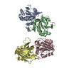
| Title | Crystal structure of the cytosolic domain of human CNNM2 in complex with AMP-PNP and Mg2+ | ||||||
 Components Components | Metal transporter CNNM2,Metal transporter CNNM2 | ||||||
 Keywords Keywords | METAL TRANSPORT / CNNM2 / Mg2+ transporter / cystathionine-beta-synthase domain / cyclic nucleotide-binding homology domain / ATP-bound / closed conformation / MEMBRANE PROTEIN | ||||||
| Function / homology |  Function and homology information Function and homology informationmagnesium ion homeostasis / magnesium ion transmembrane transporter activity / basolateral plasma membrane / glutamatergic synapse / ATP binding / plasma membrane Similarity search - Function | ||||||
| Biological species |  Homo sapiens (human) Homo sapiens (human) | ||||||
| Method |  X-RAY DIFFRACTION / SYNCHROTRON / MOLECULAR REPLACEMENT / Resolution: 3.5 Å X-RAY DIFFRACTION / SYNCHROTRON / MOLECULAR REPLACEMENT / Resolution: 3.5 Å | ||||||
 Authors Authors | Chen, Y.S. / Gehring, K. | ||||||
 Citation Citation | Journal: Structure / Year: 2020 Title: Mg2+-ATP Sensing in CNNM, a Putative Magnesium Transporter. Authors: Chen, Y.S. / Kozlov, G. / Fakih, R. / Yang, M. / Zhang, Z. / Kovrigin, E.L. / Gehring, K. | ||||||
| History |
|
- Structure visualization
| Structure viewer | Molecule: MolmilJmol/JSmol |
|---|
- Downloads & links
-Download
| PDBx/mmCIF format | 6n7e.cif.gz | 684.4 KB | Display | PDBx/mmCIF format |
|---|---|---|---|---|
| PDB format | pdb6n7e.ent.gz | 569.6 KB | Display | PDB format |
| PDBx/mmJSON format | 6n7e.json.gz | Tree view | PDBx/mmJSON format | |
| Others |  Other downloads Other downloads |
-Validation report
| Arichive directory | https://data.pdbj.org/pub/pdb/validation_reports/n7/6n7eftp://data.pdbj.org/pub/pdb/validation_reports/n7/6n7e | HTTPS FTP |
|---|
-Related structure data
| Related structure data |  6mn6C 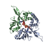 4p1oS 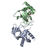 6dj3S C: citing same article ( S: Starting model for refinement |
|---|---|
| Similar structure data |


-Links
 PDBj
PDBj


- Assembly
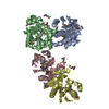
| Deposited unit | 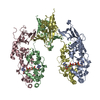
| ||||||||
|---|---|---|---|---|---|---|---|---|---|
| 1 | 
| ||||||||
| 2 | 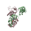
| ||||||||
| Unit cell |
|
-Components
| #1: Protein | Mass: 40802.633 Da / Num. of mol.: 4 Source method: isolated from a genetically manipulated source Source: (gene. exp.) Homo sapiens (human) / Gene: CNNM2, ACDP2 / Plasmid: pET-29a(+) / Production host:  #2: Chemical | ChemComp-ANP /   Mass: 506.196 Da / Num. of mol.: 4 / Source method: obtained synthetically / Formula: C10H17N6O12P3 / Comment: AMP-PNP, energy-carrying molecule analogue*YM Mass: 506.196 Da / Num. of mol.: 4 / Source method: obtained synthetically / Formula: C10H17N6O12P3 / Comment: AMP-PNP, energy-carrying molecule analogue*YM#3: Chemical | ChemComp-MG /   Mass: 24.305 Da / Num. of mol.: 8 / Source method: obtained synthetically / Formula: Mg Mass: 24.305 Da / Num. of mol.: 8 / Source method: obtained synthetically / Formula: Mg#4: Water | ChemComp-HOH / |  Mass: 18.015 Da / Num. of mol.: 4 / Source method: isolated from a natural source / Formula: H2O Mass: 18.015 Da / Num. of mol.: 4 / Source method: isolated from a natural source / Formula: H2O |
|---|
-Experimental details
-Experiment
| Experiment | Method: X-RAY DIFFRACTION / Number of used crystals: 1 |
|---|
- Sample preparation
| Crystal | Density Matthews: 5.03 Å3/Da / Density % sol: 75.53 % |
|---|---|
| Crystal grow | Temperature: 295 K / Method: vapor diffusion, sitting drop / pH: 6.5 Details: 0.1 M Bis-Tris, pH 6.5, 0.5 M magnesium formate, 5 mM AMP-PNP |
-Data collection
| Diffraction | Mean temperature: 100 K / Serial crystal experiment: N |
|---|---|
| Diffraction source | Source: SYNCHROTRON / Site: CLSI  / Beamline: 08ID-1 / Wavelength: 0.9796 Å / Beamline: 08ID-1 / Wavelength: 0.9796 Å |
| Detector | Type: DECTRIS PILATUS3 S 6M / Detector: PIXEL / Date: Jun 29, 2017 |
| Radiation | Protocol: SINGLE WAVELENGTH / Monochromatic (M) / Laue (L): M / Scattering type: x-ray |
| Radiation wavelength | Wavelength: 0.9796 Å / Relative weight: 1 |
| Reflection | Resolution: 3.3→50 Å / Num. obs: 36991 / % possible obs: 99.8 % / Redundancy: 3.4 % / Net I/σ(I): 17.6 |
| Reflection shell | Resolution: 3.3→3.36 Å / Redundancy: 3.4 % / Mean I/σ(I) obs: 0.5 / Num. unique obs: 683 / CC1/2: 0.148 / % possible all: 100 |
- Processing
| Software |
| ||||||||||||||||||||
|---|---|---|---|---|---|---|---|---|---|---|---|---|---|---|---|---|---|---|---|---|---|
| Refinement | Method to determine structure: MOLECULAR REPLACEMENT Starting model: 4P1O, 6DJ3 Resolution: 3.5→48.76 Å / Cross valid method: THROUGHOUT
| ||||||||||||||||||||
| Refinement step | Cycle: LAST / Resolution: 3.5→48.76 Å
|
