[English] 日本語
Yorodumi
- PDB-6n63: Crystal structure of an Iron binding protein -

+
Open data


ID or keywords:

Loading...

-
Basic information

Entry
Database: PDB / ID: 6n63
TitleCrystal structure of an Iron binding protein
ComponentsENCAPSULIN CARGO PROTEIN
KeywordsMETAL BINDING PROTEIN / IRON STORAGE / MINERALIZATION / ENCAPSULIN / FERROXIDASE
Function / homologyIMEF cargo protein / ACETATE ION / : / GLYCOLIC ACID / Uncharacterized protein
Function and homology information
Biological speciesBacillus thermotolerans (bacteria)
MethodX-RAY DIFFRACTION / SYNCHROTRON / MOLECULAR REPLACEMENT / Resolution: 1.72 Å
AuthorsBirrane, G. / Geissen, T.W.
Citation
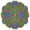
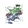
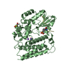
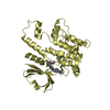
Journal: Elife / Year: 2019
Title: Large protein organelles form a new iron sequestration system with high storage capacity.
Authors: Tobias W Giessen / Benjamin J Orlando / Andrew A Verdegaal / Melissa G Chambers / Jules Gardener / David C Bell / Gabriel Birrane / Maofu Liao / Pamela A Silver /
Abstract: Iron storage proteins are essential for cellular iron homeostasis and redox balance. Ferritin proteins are the major storage units for bioavailable forms of iron. Some organisms lack ferritins, and ...Iron storage proteins are essential for cellular iron homeostasis and redox balance. Ferritin proteins are the major storage units for bioavailable forms of iron. Some organisms lack ferritins, and it is not known how they store iron. Encapsulins, a class of protein-based organelles, have recently been implicated in microbial iron and redox metabolism. Here, we report the structural and mechanistic characterization of a 42 nm two-component encapsulin-based iron storage compartment from . Using cryo-electron microscopy and x-ray crystallography, we reveal the assembly principles of a thermostable T = 4 shell topology and its catalytic ferroxidase cargo and show interactions underlying cargo-shell co-assembly. This compartment has an exceptionally large iron storage capacity storing over 23,000 iron atoms. Our results reveal a new approach for survival in diverse habitats with limited or fluctuating iron availability via an iron storage system able to store 10 to 20 times more iron than ferritin.
History
DepositionNov 24, 2018Deposition site: RCSB / Processing site: RCSB
Revision 1.0Jul 17, 2019Provider: repository / Type: Initial release
Revision 2.0Nov 15, 2023Group: Atomic model / Data collection ...Atomic model / Data collection / Database references / Derived calculations
Category: atom_site / chem_comp_atom ...atom_site / chem_comp_atom / chem_comp_bond / database_2 / pdbx_struct_conn_angle / struct_conn
Item: _atom_site.auth_atom_id / _atom_site.label_atom_id ..._atom_site.auth_atom_id / _atom_site.label_atom_id / _database_2.pdbx_DOI / _database_2.pdbx_database_accession / _pdbx_struct_conn_angle.ptnr1_auth_comp_id / _pdbx_struct_conn_angle.ptnr1_auth_seq_id / _pdbx_struct_conn_angle.ptnr1_label_asym_id / _pdbx_struct_conn_angle.ptnr1_label_atom_id / _pdbx_struct_conn_angle.ptnr1_label_comp_id / _pdbx_struct_conn_angle.ptnr1_label_seq_id / _pdbx_struct_conn_angle.ptnr3_auth_comp_id / _pdbx_struct_conn_angle.ptnr3_auth_seq_id / _pdbx_struct_conn_angle.ptnr3_label_asym_id / _pdbx_struct_conn_angle.ptnr3_label_atom_id / _pdbx_struct_conn_angle.ptnr3_label_comp_id / _pdbx_struct_conn_angle.ptnr3_label_seq_id / _pdbx_struct_conn_angle.value / _struct_conn.pdbx_dist_value / _struct_conn.ptnr1_auth_comp_id / _struct_conn.ptnr1_auth_seq_id / _struct_conn.ptnr1_label_asym_id / _struct_conn.ptnr1_label_atom_id / _struct_conn.ptnr1_label_comp_id / _struct_conn.ptnr1_label_seq_id / _struct_conn.ptnr2_symmetry

-
Structure visualization

Structure viewerMolecule:
MolmilJmol/JSmol

Downloads & links

-
Assembly

Deposited unit
A: ENCAPSULIN CARGO PROTEIN
hetero molecules


Theoretical massNumber of molelcules
Total (without water)23,6624
Polymers23,4711
Non-polymers1913
Water1,22568
1
A: ENCAPSULIN CARGO PROTEIN
hetero molecules

A: ENCAPSULIN CARGO PROTEIN
hetero molecules


Theoretical massNumber of molelcules
Total (without water)47,3248
Polymers46,9422
Non-polymers3826
Water362
TypeNameSymmetry operationNumber
identity operation1_555x,y,z1
crystal symmetry operation7_555y,x,-z1
Buried area3310 Å2
ΔGint-33 kcal/mol
Surface area14990 Å2
MethodPISA
Unit cell
Length a, b, c (Å)81.390, 81.390, 65.861
Angle α, β, γ (deg.)90.00, 90.00, 90.00
Int Tables number92
Space group name H-MP41212
Components on special symmetry positions
IDModelComponents
11A-402-

ACT

-
Components

#1: Protein ENCAPSULIN CARGO PROTEIN / IRON-MINERALIZING ENCAPSULIN-ASSOCIATED FIRMICUTE CARGO PROTEIN (IMEF)


Mass: 23471.000 Da / Num. of mol.: 1
Source method: isolated from a genetically manipulated source
Source: (gene. exp.) Bacillus thermotolerans (bacteria) / Gene: QY95_01593 / Production host: Escherichia coli (E. coli) / References: UniProt: A0A0F5HNH9
#2: Chemical ChemComp-GOA / GLYCOLIC ACID / HYDROXYACETIC ACID / HYDROXYETHANOIC ACID / Glycolic acid


Mass: 76.051 Da / Num. of mol.: 1 / Source method: obtained synthetically / Formula: C2H4O3
#3: Chemical ChemComp-ACT / ACETATE ION / Acetate


Mass: 59.044 Da / Num. of mol.: 1 / Source method: obtained synthetically / Formula: C2H3O2
#4: Chemical ChemComp-FE / FE (III) ION / Iron


Mass: 55.845 Da / Num. of mol.: 1 / Source method: obtained synthetically / Formula: Fe
#5: Water ChemComp-HOH / water / Water


Mass: 18.015 Da / Num. of mol.: 68 / Source method: isolated from a natural source / Formula: H2O

-
Experimental details

-
Experiment

ExperimentMethod: X-RAY DIFFRACTION / Number of used crystals: 1

-
Sample preparation

CrystalDensity Matthews: 2.32 Å3/Da / Density % sol: 47.06 %
Crystal growTemperature: 277 K / Method: vapor diffusion, sitting drop
Details: 10% v/v Pentaerythritol ethoxylate (3/4 EO/OH) and 10% butanol
PH range: 7.5

-
Data collection

DiffractionMean temperature: 100 K / Serial crystal experiment: N
Diffraction sourceSource: SYNCHROTRON / Site: ESRF / Beamline: ID30B / Wavelength: 0.9762 Å
DetectorType: DECTRIS PILATUS3 6M / Detector: PIXEL / Date: Sep 5, 2018
RadiationProtocol: SINGLE WAVELENGTH / Monochromatic (M) / Laue (L): M / Scattering type: x-ray
Radiation wavelengthWavelength: 0.9762 Å / Relative weight: 1
ReflectionResolution: 1.719→58 Å / Num. obs: 24131 / % possible obs: 99.7 % / Redundancy: 9.1 % / CC1/2: 0.982 / Rpim(I) all: 0.026 / Net I/σ(I): 29
Reflection shellResolution: 1.72→1.78 Å / Redundancy: 8.1 % / Mean I/σ(I) obs: 1 / Num. unique obs: 2353 / CC1/2: 0.681 / Rpim(I) all: 0.504 / % possible all: 99.7

-
Processing

Software
NameVersionClassification
PHENIX1.14_3260refinement
XDSdata reduction
XDSdata scaling
Arcimboldophasing
RefinementMethod to determine structure: MOLECULAR REPLACEMENT
Starting model: in-house model

Resolution: 1.72→40.7 Å / SU ML: 0.23 / Cross valid method: FREE R-VALUE / σ(F): 1.34 / Phase error: 26.87
RfactorNum. reflection% reflectionSelection details
Rfree0.216 1164 4.84 %Random selection
Rwork0.193 ---
obs0.194 24065 99.8 %-
Solvent computationShrinkage radii: 0.9 Å / VDW probe radii: 1.11 Å
Refinement stepCycle: LAST / Resolution: 1.72→40.7 Å
ProteinNucleic acidLigandSolventTotal
Num. atoms1191 0 10 68 1269
Refine LS restraints
Refine-IDTypeDev idealNumber
X-RAY DIFFRACTIONf_bond_d0.021229
X-RAY DIFFRACTIONf_angle_d1.361656
X-RAY DIFFRACTIONf_dihedral_angle_d6.1091048
X-RAY DIFFRACTIONf_chiral_restr0.086177
X-RAY DIFFRACTIONf_plane_restr0.01221
LS refinement shell
Resolution (Å)Rfactor RfreeNum. reflection RfreeRfactor RworkNum. reflection RworkRefine-ID% reflection obs (%)
1.7194-1.79760.3081520.31272772X-RAY DIFFRACTION99
1.7976-1.89240.32091310.2782839X-RAY DIFFRACTION100
1.8924-2.0110.2481600.24882802X-RAY DIFFRACTION100
2.011-2.16620.25331270.21352824X-RAY DIFFRACTION100
2.1662-2.38420.2031330.1952864X-RAY DIFFRACTION100
2.3842-2.72910.22121470.1852867X-RAY DIFFRACTION100
2.7291-3.43820.21551610.19752879X-RAY DIFFRACTION100
3.4382-40.70640.20131530.17523054X-RAY DIFFRACTION100

+
About Yorodumi

-
News

-
Feb 9, 2022. New format data for meta-information of EMDB entries

New format data for meta-information of EMDB entries

  • Version 3 of the EMDB header file is now the official format.
  • The previous official version 1.9 will be removed from the archive.

Related info.:EMDB header

External links:wwPDB to switch to version 3 of the EMDB data model

-
Aug 12, 2020. Covid-19 info

Covid-19 info

URL: https://pdbj.org/emnavi/covid19.php

New page: Covid-19 featured information page in EM Navigator.

Related info.:Covid-19 info / Mar 5, 2020. Novel coronavirus structure data

+
Mar 5, 2020. Novel coronavirus structure data

Novel coronavirus structure data

Related info.:Yorodumi Speices / Aug 12, 2020. Covid-19 info

External links:COVID-19 featured content - PDBj / Molecule of the Month (242):Coronavirus Proteases

+
Jan 31, 2019. EMDB accession codes are about to change! (news from PDBe EMDB page)

EMDB accession codes are about to change! (news from PDBe EMDB page)

  • The allocation of 4 digits for EMDB accession codes will soon come to an end. Whilst these codes will remain in use, new EMDB accession codes will include an additional digit and will expand incrementally as the available range of codes is exhausted. The current 4-digit format prefixed with “EMD-” (i.e. EMD-XXXX) will advance to a 5-digit format (i.e. EMD-XXXXX), and so on. It is currently estimated that the 4-digit codes will be depleted around Spring 2019, at which point the 5-digit format will come into force.
  • The EM Navigator/Yorodumi systems omit the EMD- prefix.

Related info.:Q: What is EMD? / ID/Accession-code notation in Yorodumi/EM Navigator

External links:EMDB Accession Codes are Changing Soon! / Contact to PDBj

+
Jul 12, 2017. Major update of PDB

Major update of PDB

  • wwPDB released updated PDB data conforming to the new PDBx/mmCIF dictionary.
  • This is a major update changing the version number from 4 to 5, and with Remediation, in which all the entries are updated.
  • In this update, many items about electron microscopy experimental information are reorganized (e.g. em_software).
  • Now, EM Navigator and Yorodumi are based on the updated data.

External links:wwPDB Remediation / Enriched Model Files Conforming to OneDep Data Standards Now Available in the PDB FTP Archive

-
Yorodumi

Thousand views of thousand structures

  • Yorodumi is a browser for structure data from EMDB, PDB, SASBDB, etc.
  • This page is also the successor to EM Navigator detail page, and also detail information page/front-end page for Omokage search.
  • The word "yorodu" (or yorozu) is an old Japanese word meaning "ten thousand". "mi" (miru) is to see.

Related info.:EMDB / PDB / SASBDB / Comparison of 3 databanks / Yorodumi Search / Aug 31, 2016. New EM Navigator & Yorodumi / Yorodumi Papers / Jmol/JSmol / Function and homology information / Changes in new EM Navigator and Yorodumi

Read more