Mass: 37298.281 Da / Num. of mol.: 1 Source method: isolated from a genetically manipulated source Source: (gene. exp.) Influenza A virus / Strain: A/equine/New York/49/1973(H7N7) / Gene: HA / Production host: Trichoplusia ni (cabbage looper) / References: UniProt: A0A348FV55
#2: Protein
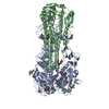
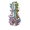
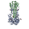
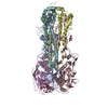
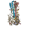
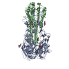
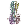
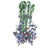
HEMAGGLUTININHA2SUBUNIT
Mass: 21533.803 Da / Num. of mol.: 1 Source method: isolated from a genetically manipulated source Source: (gene. exp.) Influenza A virus / Strain: A/equine/New York/49/1973(H7N7) / Gene: HA / Production host: Trichoplusia ni (cabbage looper) / References: UniProt: A0A348FV55
Resolution: 3.3→46.24 Å / Cor.coef. Fo:Fc: 0.895 / Cor.coef. Fo:Fc free: 0.869 / SU B: 16.434 / SU ML: 0.264 / Cross valid method: THROUGHOUT / ESU R: 0.87 / ESU R Free: 0.372 / Details: HYDROGENS HAVE BEEN ADDED IN THE RIDING POSITIONS
Rfactor
Num. reflection
% reflection
Selection details
Rfree
0.23738
986
5.1 %
RANDOM
Rwork
0.19997
-
-
-
obs
0.20181
18177
99.71 %
-
Solvent computation
Ion probe radii: 0.8 Å / Shrinkage radii: 0.8 Å / VDW probe radii: 1.2 Å
 Movie
Movie Controller

 Yorodumi
 Open data
 Basic information
 Components
 Keywords
 Function and homology information
 Influenza A virus
 X-RAY DIFFRACTION /
  Authors
 United States, 1items
  Citation
 Structure visualization
 Downloads & links
 Other downloads
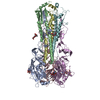

 PDBj
PDBj


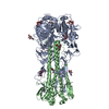
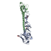
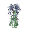
 Assembly
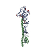
 Trichoplusia ni (cabbage looper) / References: UniProt: A0A348FV55
 Type: D-saccharide, beta linking / Mass: 221.208 Da / Num. of mol.: 2


 Mass: 150.173 Da / Num. of mol.: 1 / Source method: obtained synthetically / Formula: C6H14O4
 Mass: 106.120 Da / Num. of mol.: 1 / Source method: obtained synthetically / Formula: C4H10O3
 Mass: 62.068 Da / Num. of mol.: 3 / Source method: obtained synthetically / Formula: C2H6O2
 Mass: 194.226 Da / Num. of mol.: 1 / Source method: obtained synthetically / Formula: C8H18O5 / Comment: precipitant*YM
 Mass: 96.063 Da / Num. of mol.: 2 / Source method: obtained synthetically / Formula: SO4
 Sample preparation
 Processing