 Movie
Movie Controller


+ Open data

- Basic information
| Entry | Database: PDB / ID: 6mvm | ||||||||||||
|---|---|---|---|---|---|---|---|---|---|---|---|---|---|
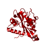
| Title | LasR LBD L130F:3OC14HSL complex | ||||||||||||
 Components Components | Transcriptional regulator LasR | ||||||||||||
 Keywords Keywords | TRANSCRIPTION / transcriptional activator protein / 3-Oxo-N-(2-oxooxolan-3-yl)tetradecanamide | ||||||||||||
| Function / homology |  Function and homology information Function and homology information | ||||||||||||
| Biological species |   Pseudomonas aeruginosa (bacteria) Pseudomonas aeruginosa (bacteria) | ||||||||||||
| Method |  X-RAY DIFFRACTION / MOLECULAR REPLACEMENT / Resolution: 1.895 Å X-RAY DIFFRACTION / MOLECULAR REPLACEMENT / Resolution: 1.895 Å | ||||||||||||
 Authors Authors | Paczkowski, J.E. / Bassler, B.L. | ||||||||||||
| Funding support |  United States, 3items United States, 3items
| ||||||||||||
 Citation Citation | Journal: Acs Chem.Biol. / Year: 2019 Title: An Autoinducer Analogue Reveals an Alternative Mode of Ligand Binding for the LasR Quorum-Sensing Receptor. Authors: Paczkowski, J.E. / McCready, A.R. / Cong, J.P. / Li, Z. / Jeffrey, P.D. / Smith, C.D. / Henke, B.R. / Hughson, F.M. / Bassler, B.L. #1: Journal: Proc. Natl. Acad. Sci. U.S.A. / Year: 2019Title: Structural determinants driving homoserine lactone ligand selection in thePseudomonas aeruginosaLasR quorum-sensing receptor. Authors: McCready, A.R. / Paczkowski, J.E. / Henke, B.R. / Bassler, B.L. | ||||||||||||
| History |
|
- Structure visualization
| Structure viewer | Molecule: MolmilJmol/JSmol |
|---|
- Downloads & links
-Download
| PDBx/mmCIF format | 6mvm.cif.gz | 82.2 KB | Display | PDBx/mmCIF format |
|---|---|---|---|---|
| PDB format | pdb6mvm.ent.gz | 60.4 KB | Display | PDB format |
| PDBx/mmJSON format | 6mvm.json.gz | Tree view | PDBx/mmJSON format | |
| Others |  Other downloads Other downloads |
-Validation report
| Arichive directory | https://data.pdbj.org/pub/pdb/validation_reports/mv/6mvmftp://data.pdbj.org/pub/pdb/validation_reports/mv/6mvm | HTTPS FTP |
|---|
-Related structure data
| Related structure data | 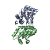 6mwhC 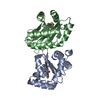 6mwlC 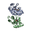 6mwwC 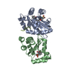 6mwzC  2uv0S S: Starting model for refinement C: citing same article ( |
|---|---|
| Similar structure data |


-Links
 PDBj
PDBj- Assembly
| Deposited unit | 
| ||||||||
|---|---|---|---|---|---|---|---|---|---|
| 1 |
| ||||||||
| Unit cell |
|
-Components
| #1: Protein | Mass: 18220.516 Da / Num. of mol.: 2 / Fragment: UNP residues 7-168 / Mutation: L130F Source method: isolated from a genetically manipulated source Source: (gene. exp.) Pseudomonas aeruginosa (strain UCBPP-PA14) (bacteria)Strain: UCBPP-PA14 / Gene: lasR, PA14_45960 / Production host: #2: Chemical |   Mass: 325.443 Da / Num. of mol.: 2 / Source method: obtained synthetically / Formula: C18H31NO4 / Feature type: SUBJECT OF INVESTIGATION Mass: 325.443 Da / Num. of mol.: 2 / Source method: obtained synthetically / Formula: C18H31NO4 / Feature type: SUBJECT OF INVESTIGATION#3: Water | ChemComp-HOH / |  Mass: 18.015 Da / Num. of mol.: 127 / Source method: isolated from a natural source / Formula: H2O Mass: 18.015 Da / Num. of mol.: 127 / Source method: isolated from a natural source / Formula: H2O |
|---|
-Experimental details
-Experiment
| Experiment | Method: X-RAY DIFFRACTION / Number of used crystals: 1 |
|---|
- Sample preparation
| Crystal | Density Matthews: 2.35 Å3/Da / Density % sol: 47.56 % |
|---|---|
| Crystal grow | Temperature: 298 K / Method: vapor diffusion, hanging drop / Details: 200 mM magnesium nitrate, 20% PEG3350 |
-Data collection
| Diffraction | Mean temperature: 100 K / Serial crystal experiment: N |
|---|---|
| Diffraction source | Source: ROTATING ANODE / Type: RIGAKU RUH3R / Wavelength: 1 Å |
| Detector | Type: RIGAKU RAXIS IV++ / Detector: IMAGE PLATE / Date: Dec 7, 2017 |
| Radiation | Protocol: SINGLE WAVELENGTH / Monochromatic (M) / Laue (L): M / Scattering type: x-ray |
| Radiation wavelength | Wavelength: 1 Å / Relative weight: 1 |
| Reflection | Resolution: 1.895→30 Å / Num. obs: 25887 / % possible obs: 96.6 % / Redundancy: 2.5 % / Net I/σ(I): 14.4 |
| Reflection shell | Resolution: 1.9→1.98 Å |
- Processing
| Software |
| ||||||||||||||||||||||||||||||||||||||||||||||||||||||||||||||||||||||
|---|---|---|---|---|---|---|---|---|---|---|---|---|---|---|---|---|---|---|---|---|---|---|---|---|---|---|---|---|---|---|---|---|---|---|---|---|---|---|---|---|---|---|---|---|---|---|---|---|---|---|---|---|---|---|---|---|---|---|---|---|---|---|---|---|---|---|---|---|---|---|---|
| Refinement | Method to determine structure: MOLECULAR REPLACEMENT Starting model: PDB entry 2UV0 Resolution: 1.895→29.568 Å / Cross valid method: FREE R-VALUE / σ(F): 0 / Phase error: 31.58
| ||||||||||||||||||||||||||||||||||||||||||||||||||||||||||||||||||||||
| Solvent computation | Shrinkage radii: 0.9 Å / VDW probe radii: 1.11 Å | ||||||||||||||||||||||||||||||||||||||||||||||||||||||||||||||||||||||
| Refinement step | Cycle: LAST / Resolution: 1.895→29.568 Å
| ||||||||||||||||||||||||||||||||||||||||||||||||||||||||||||||||||||||
| Refine LS restraints |
| ||||||||||||||||||||||||||||||||||||||||||||||||||||||||||||||||||||||
| LS refinement shell |
|
