 Movie
Movie Controller

[English] 日本語
 Yorodumi
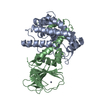
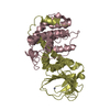
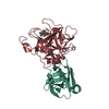
- PDB-2z2s: Crystal Structure of Rhodobacter sphaeroides SigE in complex with... -
+ Open data

- Basic information
| Entry | Database: PDB / ID: 2z2s | ||||||
|---|---|---|---|---|---|---|---|
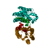
| Title | Crystal Structure of Rhodobacter sphaeroides SigE in complex with the anti-sigma ChrR | ||||||
 Components Components |
| ||||||
 Keywords Keywords | TRANSCRIPTION / ECF sigma factor / anti-sigma factor / cupin fold / DNA-binding / Transcription regulation / Activator / Metal-binding / Zinc binding transcription factor | ||||||
| Function / homology |  Function and homology information Function and homology informationsigma factor activity / DNA-templated transcription initiation / DNA-templated transcription / DNA binding / metal ion binding Similarity search - Function | ||||||
| Biological species |  Rhodobacter sphaeroides (bacteria) Rhodobacter sphaeroides (bacteria) | ||||||
| Method |  X-RAY DIFFRACTION / SYNCHROTRON / MOLECULAR REPLACEMENT / Resolution: 2.7 Å X-RAY DIFFRACTION / SYNCHROTRON / MOLECULAR REPLACEMENT / Resolution: 2.7 Å | ||||||
 Authors Authors | Darst, S.A. / Campbell, E.A. | ||||||
 Citation Citation | Journal: Mol.Cell / Year: 2007 Title: A conserved structural module regulates transcriptional responses to diverse stress signals in bacteria. Authors: Campbell, E.A. / Greenwell, R. / Anthony, J.R. / Wang, S. / Lim, L. / Das, K. / Sofia, H.J. / Donohue, T.J. / Darst, S.A. | ||||||
| History |
|
- Structure visualization
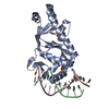
| Structure viewer | Molecule: MolmilJmol/JSmol |
|---|
- Downloads & links
-Download
| PDBx/mmCIF format | 2z2s.cif.gz | 271.3 KB | Display | PDBx/mmCIF format |
|---|---|---|---|---|
| PDB format | pdb2z2s.ent.gz | 218 KB | Display | PDB format |
| PDBx/mmJSON format | 2z2s.json.gz | Tree view | PDBx/mmJSON format | |
| Others |  Other downloads Other downloads |
-Validation report
| Arichive directory | https://data.pdbj.org/pub/pdb/validation_reports/z2/2z2sftp://data.pdbj.org/pub/pdb/validation_reports/z2/2z2s | HTTPS FTP |
|---|
-Related structure data


-Links
 PDBj
PDBj
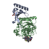
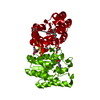
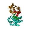
- Assembly
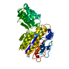
| Deposited unit | 
| ||||||||
|---|---|---|---|---|---|---|---|---|---|
| 1 | 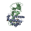
| ||||||||
| 2 | 
| ||||||||
| 3 | 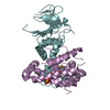
| ||||||||
| 4 | 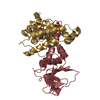
| ||||||||
| Unit cell |
|
-Components
| #1: Protein | Mass: 21309.201 Da / Num. of mol.: 4 Source method: isolated from a genetically manipulated source Source: (gene. exp.) Rhodobacter sphaeroides (bacteria) / Strain: 2.4.1 / Gene: rpoE / Plasmid: pET28a derivitive / Species (production host): Escherichia coli / Production host: #2: Protein | Mass: 21804.225 Da / Num. of mol.: 4 Source method: isolated from a genetically manipulated source Source: (gene. exp.) Rhodobacter sphaeroides (bacteria) / Strain: 2.4.1 / Gene: chrR / Plasmid: pET28a derivitive / Species (production host): Escherichia coli / Production host: #3: Chemical | ChemComp-ZN /   Mass: 65.409 Da / Num. of mol.: 4 / Source method: obtained synthetically / Formula: Zn Mass: 65.409 Da / Num. of mol.: 4 / Source method: obtained synthetically / Formula: Zn#4: Chemical |   Mass: 96.063 Da / Num. of mol.: 2 / Source method: obtained synthetically / Formula: SO4 Mass: 96.063 Da / Num. of mol.: 2 / Source method: obtained synthetically / Formula: SO4#5: Water | ChemComp-HOH / |  Mass: 18.015 Da / Num. of mol.: 381 / Source method: isolated from a natural source / Formula: H2O Mass: 18.015 Da / Num. of mol.: 381 / Source method: isolated from a natural source / Formula: H2OHas protein modification | Y | |
|---|
-Experimental details
-Experiment
| Experiment | Method: X-RAY DIFFRACTION / Number of used crystals: 1 |
|---|
- Sample preparation
| Crystal | Density Matthews: 2.27 Å3/Da / Density % sol: 45.88 % |
|---|---|
| Crystal grow | Temperature: 295.15 K / Method: vapor diffusion, hanging drop / pH: 6.5 Details: 0.1 M MES, 0.2 M ammonium sulfate, 14-18% polyethylene glycol monomethylether 5000 (PEG5KMME), pH 6.5, VAPOR DIFFUSION, HANGING DROP, temperature 295.15K |
-Data collection
| Diffraction | Mean temperature: 173 K |
|---|---|
| Diffraction source | Source: SYNCHROTRON / Site: APS  / Beamline: 19-ID / Wavelength: 0.97969 Å / Beamline: 19-ID / Wavelength: 0.97969 Å |
| Detector | Type: ADSC QUANTUM 315 / Detector: CCD / Date: Feb 3, 2003 |
| Radiation | Monochromator: Si 111 channel / Protocol: SINGLE WAVELENGTH / Monochromatic (M) / Laue (L): M / Scattering type: x-ray |
| Radiation wavelength | Wavelength: 0.97969 Å / Relative weight: 1 |
| Reflection | Resolution: 2.6→25 Å / Num. obs: 24975 / % possible obs: 75.8 % / Observed criterion σ(F): 0 / Observed criterion σ(I): 0 / Redundancy: 2.4 % / Rmerge(I) obs: 0.079 / Rsym value: 0.079 / Net I/σ(I): 9.8 |
| Reflection shell | Resolution: 2.6→2.69 Å / Redundancy: 2.1 % / Rmerge(I) obs: 0.079 / Mean I/σ(I) obs: 2.14 / Num. unique all: 2887 / Rsym value: 0.079 / % possible all: 60.7 |
- Processing
| Software |
| |||||||||||||||||||||||||
|---|---|---|---|---|---|---|---|---|---|---|---|---|---|---|---|---|---|---|---|---|---|---|---|---|---|---|
| Refinement | Method to determine structure: MOLECULAR REPLACEMENT / Resolution: 2.7→25 Å / Cross valid method: THROUGHOUT / Stereochemistry target values: maximum likelihood
| |||||||||||||||||||||||||
| Refinement step | Cycle: LAST / Resolution: 2.7→25 Å
| |||||||||||||||||||||||||
| Refine LS restraints |
|
