 Movie
Movie Controller

[English] 日本語
 Yorodumi
- PDB-6mvf: Crystal structure of FMN-binding beta-glucuronidase from Facaelib... -
+ Open data

- Basic information
| Entry | Database: PDB / ID: 6mvf | ||||||
|---|---|---|---|---|---|---|---|
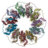
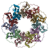
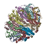
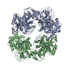
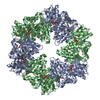
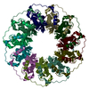
| Title | Crystal structure of FMN-binding beta-glucuronidase from Facaelibacterium prausnitzii L2-6 | ||||||
 Components Components | Beta-galactosidase/beta-glucuronidase | ||||||
 Keywords Keywords | HYDROLASE / glycoside hydrolase / FMN / beta-glucuronidase | ||||||
| Function / homology |  Function and homology information Function and homology informationbeta-galactosidase / beta-galactosidase activity / cellulose catabolic process / nucleotide binding Similarity search - Function | ||||||
| Biological species |  Faecalibacterium prausnitzii L2-6 (bacteria) Faecalibacterium prausnitzii L2-6 (bacteria) | ||||||
| Method |  X-RAY DIFFRACTION / SYNCHROTRON / MOLECULAR REPLACEMENT / Resolution: 2.55 Å X-RAY DIFFRACTION / SYNCHROTRON / MOLECULAR REPLACEMENT / Resolution: 2.55 Å | ||||||
 Authors Authors | Pellock, S.J. / Redinbo, M.R. | ||||||
| Funding support |  United States, 1items United States, 1items
| ||||||
 Citation Citation | Journal: J. Mol. Biol. / Year: 2019 Title: Discovery and Characterization of FMN-Binding beta-Glucuronidases in the Human Gut Microbiome. Authors: Pellock, S.J. / Walton, W.G. / Ervin, S.M. / Torres-Rivera, D. / Creekmore, B.C. / Bergan, G. / Dunn, Z.D. / Li, B. / Tripathy, A. / Redinbo, M.R. | ||||||
| History |
|
- Structure visualization
| Structure viewer | Molecule: MolmilJmol/JSmol |
|---|
- Downloads & links
-Download
| PDBx/mmCIF format | 6mvf.cif.gz | 783 KB | Display | PDBx/mmCIF format |
|---|---|---|---|---|
| PDB format | pdb6mvf.ent.gz | 637.7 KB | Display | PDB format |
| PDBx/mmJSON format | 6mvf.json.gz | Tree view | PDBx/mmJSON format | |
| Others |  Other downloads Other downloads |
-Validation report
| Arichive directory | https://data.pdbj.org/pub/pdb/validation_reports/mv/6mvfftp://data.pdbj.org/pub/pdb/validation_reports/mv/6mvf | HTTPS FTP |
|---|
-Related structure data
| Related structure data |  6mvgC 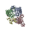 6mvhC 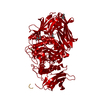 3cmgS S: Starting model for refinement C: citing same article ( |
|---|---|
| Similar structure data |


-Links
 PDBj
PDBj

- Assembly
| Deposited unit | 
| ||||||||
|---|---|---|---|---|---|---|---|---|---|
| 1 | 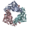
| ||||||||
| 2 | 
| ||||||||
| Unit cell |
|
-Components
| #1: Protein | Mass: 86281.453 Da / Num. of mol.: 6 Source method: isolated from a genetically manipulated source Source: (gene. exp.) Faecalibacterium prausnitzii L2-6 (bacteria)Gene: FP2_04120 / Production host: #2: Chemical | ChemComp-FMN /   Mass: 456.344 Da / Num. of mol.: 6 / Source method: isolated from a natural source / Formula: C17H21N4O9P / Feature type: SUBJECT OF INVESTIGATION Mass: 456.344 Da / Num. of mol.: 6 / Source method: isolated from a natural source / Formula: C17H21N4O9P / Feature type: SUBJECT OF INVESTIGATION#3: Water | ChemComp-HOH / |  Mass: 18.015 Da / Num. of mol.: 1398 / Source method: isolated from a natural source / Formula: H2O Mass: 18.015 Da / Num. of mol.: 1398 / Source method: isolated from a natural source / Formula: H2O |
|---|
-Experimental details
-Experiment
| Experiment | Method: X-RAY DIFFRACTION / Number of used crystals: 1 |
|---|
- Sample preparation
| Crystal | Density Matthews: 2.46 Å3/Da / Density % sol: 49.94 % |
|---|---|
| Crystal grow | Temperature: 293 K / Method: vapor diffusion, sitting drop Details: 11.1 mg/mL protein, 0.2 M magnesium chloride, 10% w/v PEG 3000, 0.1 M sodium cacodylate |
-Data collection
| Diffraction | Mean temperature: 100 K / Serial crystal experiment: N |
|---|---|
| Diffraction source | Source: SYNCHROTRON / Site: APS / Beamline: 23-ID-D / Wavelength: 1 Å |
| Detector | Type: DECTRIS PILATUS3 6M / Detector: PIXEL / Date: Aug 10, 2017 |
| Radiation | Protocol: SINGLE WAVELENGTH / Monochromatic (M) / Laue (L): M / Scattering type: x-ray |
| Radiation wavelength | Wavelength: 1 Å / Relative weight: 1 |
| Reflection | Resolution: 2.55→29.52 Å / Num. obs: 163078 / % possible obs: 99.58 % / Redundancy: 5 % / Rmerge(I) obs: 0.1212 / Net I/σ(I): 11.22 |
| Reflection shell | Resolution: 2.55→2.641 Å / Redundancy: 4.4 % / Rmerge(I) obs: 0.5703 / Mean I/σ(I) obs: 2.58 / Num. unique obs: 16164 / % possible all: 99.53 |
- Processing
| Software |
| |||||||||||||||||||||||||||||||||||||||||||||||||||||||||||||||||||||||||||||||||||||||||||||||||||||||||
|---|---|---|---|---|---|---|---|---|---|---|---|---|---|---|---|---|---|---|---|---|---|---|---|---|---|---|---|---|---|---|---|---|---|---|---|---|---|---|---|---|---|---|---|---|---|---|---|---|---|---|---|---|---|---|---|---|---|---|---|---|---|---|---|---|---|---|---|---|---|---|---|---|---|---|---|---|---|---|---|---|---|---|---|---|---|---|---|---|---|---|---|---|---|---|---|---|---|---|---|---|---|---|---|---|---|---|
| Refinement | Method to determine structure: MOLECULAR REPLACEMENT Starting model: 3CMG Resolution: 2.55→29.524 Å / SU ML: 0.32 / Cross valid method: FREE R-VALUE / σ(F): 1.34 / Phase error: 27.48 / Stereochemistry target values: ML
| |||||||||||||||||||||||||||||||||||||||||||||||||||||||||||||||||||||||||||||||||||||||||||||||||||||||||
| Solvent computation | Shrinkage radii: 0.9 Å / VDW probe radii: 1.11 Å / Solvent model: FLAT BULK SOLVENT MODEL | |||||||||||||||||||||||||||||||||||||||||||||||||||||||||||||||||||||||||||||||||||||||||||||||||||||||||
| Refinement step | Cycle: LAST / Resolution: 2.55→29.524 Å
| |||||||||||||||||||||||||||||||||||||||||||||||||||||||||||||||||||||||||||||||||||||||||||||||||||||||||
| Refine LS restraints |
| |||||||||||||||||||||||||||||||||||||||||||||||||||||||||||||||||||||||||||||||||||||||||||||||||||||||||
| LS refinement shell |
|
