 Movie
Movie Controller

[English] 日本語
 Yorodumi
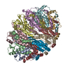
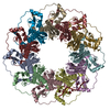
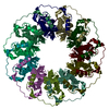
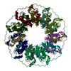
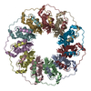
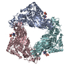
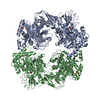
- PDB-5xrz: Structure of a ssDNA bound to the inner DNA binding site of RAD52 -
+ Open data

- Basic information
| Entry | Database: PDB / ID: 5xrz | |||||||||||||||
|---|---|---|---|---|---|---|---|---|---|---|---|---|---|---|---|---|
| Title | Structure of a ssDNA bound to the inner DNA binding site of RAD52 | |||||||||||||||
 Components Components |
| |||||||||||||||
 Keywords Keywords | RECOMBINATION / Protein-DNA complex / ssDNA annealing protein / DNA repair protein | |||||||||||||||
| Function / homology |  Function and homology information Function and homology informationdouble-strand break repair via single-strand annealing / DNA double-strand break processing involved in repair via single-strand annealing / DNA recombinase assembly / mitotic recombination / HDR through MMEJ (alt-NHEJ) / regulation of nucleotide-excision repair / HDR through Single Strand Annealing (SSA) / SUMOylation of DNA damage response and repair proteins / protein-DNA complex / double-strand break repair via homologous recombination ...double-strand break repair via single-strand annealing / DNA double-strand break processing involved in repair via single-strand annealing / DNA recombinase assembly / mitotic recombination / HDR through MMEJ (alt-NHEJ) / regulation of nucleotide-excision repair / HDR through Single Strand Annealing (SSA) / SUMOylation of DNA damage response and repair proteins / protein-DNA complex / double-strand break repair via homologous recombination / double-strand break repair / single-stranded DNA binding / cellular response to oxidative stress / DNA recombination / protein-containing complex / DNA binding / nucleoplasm / identical protein binding / nucleus Similarity search - Function | |||||||||||||||
| Biological species |  Homo sapiens (human) Homo sapiens (human)synthetic construct (others) | |||||||||||||||
| Method |  X-RAY DIFFRACTION / SYNCHROTRON / MOLECULAR REPLACEMENT / Resolution: 3.6 Å X-RAY DIFFRACTION / SYNCHROTRON / MOLECULAR REPLACEMENT / Resolution: 3.6 Å | |||||||||||||||
 Authors Authors | Saotome, M. / Saito, K. / Yasuda, T. / Sugiyama, S. / Kurumizaka, H. / Kagawa, W. | |||||||||||||||
| Funding support |  Japan, 4items Japan, 4items
| |||||||||||||||
 Citation Citation | Journal: iScience / Year: 2018 Title: Structural Basis of Homology-Directed DNA Repair Mediated by RAD52 Authors: Saotome, M. / Saito, K. / Yasuda, T. / Ohtomo, H. / Sugiyama, S. / Nishimura, Y. / Kurumizaka, H. / Kagawa, W. | |||||||||||||||
| History |
|
- Structure visualization
| Structure viewer | Molecule: MolmilJmol/JSmol |
|---|
- Downloads & links
-Download
| PDBx/mmCIF format | 5xrz.cif.gz | 415.9 KB | Display | PDBx/mmCIF format |
|---|---|---|---|---|
| PDB format | pdb5xrz.ent.gz | 339.8 KB | Display | PDB format |
| PDBx/mmJSON format | 5xrz.json.gz | Tree view | PDBx/mmJSON format | |
| Others |  Other downloads Other downloads |
-Validation report
| Arichive directory | https://data.pdbj.org/pub/pdb/validation_reports/xr/5xrzftp://data.pdbj.org/pub/pdb/validation_reports/xr/5xrz | HTTPS FTP |
|---|
-Related structure data
| Related structure data |  5xs0C 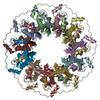 1kn0S S: Starting model for refinement C: citing same article ( |
|---|---|
| Similar structure data |

-Links
 PDBj
PDBj


- Assembly
| Deposited unit | 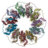
| ||||||||
|---|---|---|---|---|---|---|---|---|---|
| 1 |
| ||||||||
| Unit cell |
|
-Components
| #1: Protein | Mass: 23597.582 Da / Num. of mol.: 11 / Fragment: UNP residues 1-212 / Mutation: K102A, K133A Source method: isolated from a genetically manipulated source Source: (gene. exp.) Homo sapiens (human) / Gene: RAD52 / Plasmid: pET15b / Production host:  #2: DNA chain | | Mass: 12077.721 Da / Num. of mol.: 1 / Source method: obtained synthetically / Source: (synth.) synthetic construct (others) #3: Chemical | ChemComp-K /   Mass: 39.098 Da / Num. of mol.: 10 / Source method: obtained synthetically / Formula: K Mass: 39.098 Da / Num. of mol.: 10 / Source method: obtained synthetically / Formula: K |
|---|
-Experimental details
-Experiment
| Experiment | Method: X-RAY DIFFRACTION / Number of used crystals: 1 |
|---|
- Sample preparation
| Crystal | Density Matthews: 2.29 Å3/Da / Density % sol: 46.19 % |
|---|---|
| Crystal grow | Temperature: 293 K / Method: vapor diffusion, hanging drop / Details: calcium acetate, PEG 3,350 |
-Data collection
| Diffraction | Mean temperature: 100 K |
|---|---|
| Diffraction source | Source: SYNCHROTRON / Site: Photon Factory / Beamline: BL-1A / Wavelength: 1.1 Å |
| Detector | Type: DECTRIS PILATUS 2M-F / Detector: PIXEL / Date: Dec 7, 2014 |
| Radiation | Monochromator: Cryo-cooled channel-cut Si(111) / Protocol: SINGLE WAVELENGTH / Monochromatic (M) / Laue (L): M / Scattering type: x-ray |
| Radiation wavelength | Wavelength: 1.1 Å / Relative weight: 1 |
| Reflection | Resolution: 3.6→95.3 Å / Num. obs: 27023 / % possible obs: 96.9 % / Observed criterion σ(F): 0 / Observed criterion σ(I): 0 / Redundancy: 3.3 % / Biso Wilson estimate: 100.2 Å2 / CC1/2: 0.972 / Rmerge(I) obs: 0.103 / Rpim(I) all: 0.103 / Rrim(I) all: 0.145 / Rsym value: 0.103 / Net I/σ(I): 7.5 |
| Reflection shell | Resolution: 3.6→3.7 Å / Redundancy: 3.4 % / Rmerge(I) obs: 0.4 / Mean I/σ(I) obs: 2.7 / Num. unique obs: 4412 / CC1/2: 0.597 / Rpim(I) all: 0.4 / Rrim(I) all: 0.565 / Rsym value: 0.103 / % possible all: 97.1 |
- Processing
| Software |
| |||||||||||||||||||||||||||||||||||||||||||||||||||||||||||||||||||||||||||||
|---|---|---|---|---|---|---|---|---|---|---|---|---|---|---|---|---|---|---|---|---|---|---|---|---|---|---|---|---|---|---|---|---|---|---|---|---|---|---|---|---|---|---|---|---|---|---|---|---|---|---|---|---|---|---|---|---|---|---|---|---|---|---|---|---|---|---|---|---|---|---|---|---|---|---|---|---|---|---|
| Refinement | Method to determine structure: MOLECULAR REPLACEMENT Starting model: 1KN0 Resolution: 3.6→45.703 Å / SU ML: 0.41 / Cross valid method: THROUGHOUT / σ(F): 1.96 / Phase error: 31.2 / Stereochemistry target values: ML
| |||||||||||||||||||||||||||||||||||||||||||||||||||||||||||||||||||||||||||||
| Solvent computation | Shrinkage radii: 0.9 Å / VDW probe radii: 1.11 Å / Solvent model: FLAT BULK SOLVENT MODEL | |||||||||||||||||||||||||||||||||||||||||||||||||||||||||||||||||||||||||||||
| Refinement step | Cycle: LAST / Resolution: 3.6→45.703 Å
| |||||||||||||||||||||||||||||||||||||||||||||||||||||||||||||||||||||||||||||
| Refine LS restraints |
| |||||||||||||||||||||||||||||||||||||||||||||||||||||||||||||||||||||||||||||
| LS refinement shell |
|
