| Entry | Database: PDB / ID: 6mp2
|
|---|
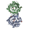
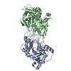
| Title | Crystal structure of BlMan5B solved by SIRAS |
|---|
 Components Components | BlMan5B |
|---|
 Keywords Keywords | HYDROLASE / Family GH5 / subfamily 18 / beta-mannosidase |
|---|
| Function / homology | Glycosidases / Glycoside hydrolase superfamily / TIM Barrel / Alpha-Beta Barrel / Alpha Beta / Glycosyl hydrolase Function and homology information Function and homology information |
|---|
| Biological species |  Bifidobacterium longum DJO10A (bacteria) Bifidobacterium longum DJO10A (bacteria) |
|---|
| Method |  X-RAY DIFFRACTION / SYNCHROTRON / MOLECULAR REPLACEMENT / SIRAS / Resolution: 1.98 Å X-RAY DIFFRACTION / SYNCHROTRON / MOLECULAR REPLACEMENT / SIRAS / Resolution: 1.98 Å |
|---|
 Authors Authors | Lorizolla-Cordeiro, R. / Giuseppe, P.O. / Murakami, M.T. |
|---|
| Funding support |  Brazil, 2items Brazil, 2items | Organization | Grant number | Country |
|---|
| Sao Paulo Research Foundation (FAPESP) | 2015/26982-0 | Brazil | | Sao Paulo Research Foundation (FAPESP) | 2016/00740-2 | Brazil |
|
|---|
 Citation Citation | Journal: J. Mol. Biol. / Year: 2019
Title: N-glycan Utilization by Bifidobacterium Gut Symbionts Involves a Specialist beta-Mannosidase.
Authors: Cordeiro, R.L. / Pirolla, R.A.S. / Persinoti, G.F. / Gozzo, F.C. / de Giuseppe, P.O. / Murakami, M.T. |
|---|
| History | | Deposition | Oct 5, 2018 | Deposition site: RCSB / Processing site: RCSB |
|---|
| Revision 1.0 | Jan 30, 2019 | Provider: repository / Type: Initial release |
|---|
| Revision 1.1 | Feb 27, 2019 | Group: Data collection / Database references / Category: citation
Item: _citation.journal_volume / _citation.page_first ..._citation.journal_volume / _citation.page_first / _citation.page_last / _citation.title |
|---|
| Revision 1.2 | Jan 1, 2020 | Group: Author supporting evidence / Category: pdbx_audit_support / Item: _pdbx_audit_support.funding_organization |
|---|
| Revision 1.3 | Mar 13, 2024 | Group: Data collection / Database references / Category: chem_comp_atom / chem_comp_bond / database_2
Item: _database_2.pdbx_DOI / _database_2.pdbx_database_accession |
|---|
|
|---|
 Movie
Movie Controller


 Open data
 Basic information
 Structure visualization
 Downloads & links
 Other downloads


 PDBj
PDBj Assembly