 Movie
Movie Controller


+ Open data

- Basic information
| Entry | Database: PDB / ID: 6mip | ||||||
|---|---|---|---|---|---|---|---|
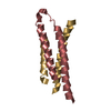
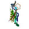
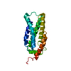
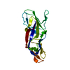
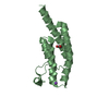
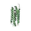
| Title | Crystal structure of Taf14 YEATS domain G82A mutant | ||||||
 Components Components | Transcription initiation factor TFIID subunit 14 | ||||||
 Keywords Keywords | TRANSCRIPTION / Epigenetic / Histone reader | ||||||
| Function / homology |  Function and homology information Function and homology informationNuA3b histone acetyltransferase complex / NuA3 histone acetyltransferase complex / NuA3a histone acetyltransferase complex / Ino80 complex / transcription factor TFIIF complex / mediator complex / SWI/SNF complex / transcription factor TFIID complex / RNA polymerase II general transcription initiation factor activity / RNA polymerase II preinitiation complex assembly ...NuA3b histone acetyltransferase complex / NuA3 histone acetyltransferase complex / NuA3a histone acetyltransferase complex / Ino80 complex / transcription factor TFIIF complex / mediator complex / SWI/SNF complex / transcription factor TFIID complex / RNA polymerase II general transcription initiation factor activity / RNA polymerase II preinitiation complex assembly / transcription initiation at RNA polymerase II promoter / histone binding / transcription by RNA polymerase II / chromatin remodeling / DNA repair / DNA-templated transcription / regulation of DNA-templated transcription / chromatin / regulation of transcription by RNA polymerase II / positive regulation of transcription by RNA polymerase II / nucleus Similarity search - Function | ||||||
| Biological species |  | ||||||
| Method |  X-RAY DIFFRACTION / MOLECULAR REPLACEMENT / Resolution: 2 Å X-RAY DIFFRACTION / MOLECULAR REPLACEMENT / Resolution: 2 Å | ||||||
 Authors Authors | Klein, B.J. / Andrews, F.H. / Kutateladze, T.G. | ||||||
 Citation Citation | Journal: Nat Commun / Year: 2018 Title: Structural insights into the pi-pi-pi stacking mechanism and DNA-binding activity of the YEATS domain. Authors: Klein, B.J. / Vann, K.R. / Andrews, F.H. / Wang, W.W. / Zhang, J. / Zhang, Y. / Beloglazkina, A.A. / Mi, W. / Li, Y. / Li, H. / Shi, X. / Kutateladze, A.G. / Strahl, B.D. / Liu, W.R. / Kutateladze, T.G. | ||||||
| History |
|
- Structure visualization
| Structure viewer | Molecule: MolmilJmol/JSmol |
|---|
- Downloads & links
-Download
| PDBx/mmCIF format | 6mip.cif.gz | 76.2 KB | Display | PDBx/mmCIF format |
|---|---|---|---|---|
| PDB format | pdb6mip.ent.gz | 55 KB | Display | PDB format |
| PDBx/mmJSON format | 6mip.json.gz | Tree view | PDBx/mmJSON format | |
| Others |  Other downloads Other downloads |
-Validation report
| Summary document | 6mip_validation.pdf.gz | 436.2 KB | Display | wwPDB validaton report |
|---|---|---|---|---|
| Full document | 6mip_full_validation.pdf.gz | 437 KB | Display | |
| Data in XML | 6mip_validation.xml.gz | 9.3 KB | Display | |
| Data in CIF | 6mip_validation.cif.gz | 12.5 KB | Display | |
| Arichive directory | https://data.pdbj.org/pub/pdb/validation_reports/mi/6mipftp://data.pdbj.org/pub/pdb/validation_reports/mi/6mip | HTTPS FTP |
-Related structure data
| Related structure data |  6milC  6mimC  6minC  6mioC 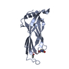 6miqC 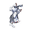 5iokS S: Starting model for refinement C: citing same article ( |
|---|---|
| Similar structure data |


-Links
 PDBj
PDBj- Assembly
| Deposited unit | 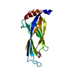
| ||||||||
|---|---|---|---|---|---|---|---|---|---|
| 1 |
| ||||||||
| Unit cell |
|
-Components
| #1: Protein | Mass: 16194.522 Da / Num. of mol.: 1 / Fragment: YEATS domain residues 1-137 / Mutation: G82A Source method: isolated from a genetically manipulated source Source: (gene. exp.) Strain: ATCC 204508 / S288c / Gene: TAF14, ANC1, CST10, SWP29, TAF30, TFG3, YPL129W / Production host:  |
|---|---|
| #2: Chemical | ChemComp-PEG /   Mass: 106.120 Da / Num. of mol.: 1 / Source method: obtained synthetically / Formula: C4H10O3 Mass: 106.120 Da / Num. of mol.: 1 / Source method: obtained synthetically / Formula: C4H10O3 |
| #3: Water | ChemComp-HOH /  Mass: 18.015 Da / Num. of mol.: 146 / Source method: isolated from a natural source / Formula: H2O Mass: 18.015 Da / Num. of mol.: 146 / Source method: isolated from a natural source / Formula: H2O |
-Experimental details
-Experiment
| Experiment | Method: X-RAY DIFFRACTION / Number of used crystals: 1 |
|---|
- Sample preparation
| Crystal | Density Matthews: 3 Å3/Da / Density % sol: 59.05 % |
|---|---|
| Crystal grow | Temperature: 298 K / Method: vapor diffusion, sitting drop / Details: 48% PEG600 with 0.2 M sodium citrate (pH 6.0) |
-Data collection
| Diffraction | Mean temperature: 100 K |
|---|---|
| Diffraction source | Source: ROTATING ANODE / Type: RIGAKU MICROMAX-007 HF / Wavelength: 1.54 Å |
| Detector | Type: DECTRIS PILATUS 200K / Detector: PIXEL / Date: Apr 22, 2016 |
| Radiation | Protocol: SINGLE WAVELENGTH / Monochromatic (M) / Laue (L): M / Scattering type: x-ray |
| Radiation wavelength | Wavelength: 1.54 Å / Relative weight: 1 |
| Reflection | Resolution: 2→50 Å / Num. obs: 17536 / % possible obs: 92.8 % / Redundancy: 3.3 % / Rsym value: 0.042 / Net I/σ(I): 35.9 |
| Reflection shell | Resolution: 2→2.03 Å / Rsym value: 0.092 |
- Processing
| Software |
| ||||||||||||||||||||||||||||||||||||||||
|---|---|---|---|---|---|---|---|---|---|---|---|---|---|---|---|---|---|---|---|---|---|---|---|---|---|---|---|---|---|---|---|---|---|---|---|---|---|---|---|---|---|
| Refinement | Method to determine structure: MOLECULAR REPLACEMENT Starting model: 5IOK Resolution: 2→37.107 Å / SU ML: 0.18 / Cross valid method: FREE R-VALUE / σ(F): 0 / Phase error: 21.94
| ||||||||||||||||||||||||||||||||||||||||
| Solvent computation | Shrinkage radii: 0.9 Å / VDW probe radii: 1.11 Å | ||||||||||||||||||||||||||||||||||||||||
| Refinement step | Cycle: LAST / Resolution: 2→37.107 Å
| ||||||||||||||||||||||||||||||||||||||||
| Refine LS restraints |
| ||||||||||||||||||||||||||||||||||||||||
| LS refinement shell |
| ||||||||||||||||||||||||||||||||||||||||
| Refinement TLS params. | Method: refined / Origin x: -43.8938 Å / Origin y: 13.2123 Å / Origin z: 2.198 Å
| ||||||||||||||||||||||||||||||||||||||||
| Refinement TLS group | Selection details: (chain 'A' and resid -1 through 137) |
