 Movie
Movie Controller

[English] 日本語
 Yorodumi
- PDB-6mg0: Crystal structure of a 5-domain construct of LgrA in the thiolati... -
+ Open data

- Basic information
| Entry | Database: PDB / ID: 6mg0 | |||||||||
|---|---|---|---|---|---|---|---|---|---|---|
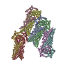
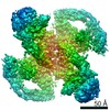
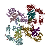
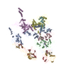
| Title | Crystal structure of a 5-domain construct of LgrA in the thiolation state | |||||||||
 Components Components | Linear gramicidin synthase subunit A | |||||||||
 Keywords Keywords | LIGASE / nonribosomal peptide synthetase / tailoring domain / NRPS / enzyme / natural product / linear gramicidin | |||||||||
| Function / homology |  Function and homology information Function and homology informationamino acid activation for nonribosomal peptide biosynthetic process / secondary metabolite biosynthetic process / lipid biosynthetic process / ligase activity / phosphopantetheine binding / antibiotic biosynthetic process / cytoplasm Similarity search - Function | |||||||||
| Biological species |  Brevibacillus parabrevis (bacteria) Brevibacillus parabrevis (bacteria) | |||||||||
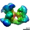
| Method |  X-RAY DIFFRACTION / SYNCHROTRON / MOLECULAR REPLACEMENT / Resolution: 6 Å X-RAY DIFFRACTION / SYNCHROTRON / MOLECULAR REPLACEMENT / Resolution: 6 Å | |||||||||
 Authors Authors | Reimer, J.M. / Eivaskhani, M. / Harb, I. / Schmeing, T.M. | |||||||||
| Funding support |  Canada, 2items Canada, 2items
| |||||||||
 Citation Citation | Journal: Science / Year: 2019 Title: Structures of a dimodular nonribosomal peptide synthetase reveal conformational flexibility. Authors: Reimer, J.M. / Eivaskhani, M. / Harb, I. / Guarne, A. / Weigt, M. / Schmeing, T.M. | |||||||||
| History |
|
- Structure visualization
| Structure viewer | Molecule: MolmilJmol/JSmol |
|---|
- Downloads & links
-Download
| PDBx/mmCIF format | 6mg0.cif.gz | 694.4 KB | Display | PDBx/mmCIF format |
|---|---|---|---|---|
| PDB format | pdb6mg0.ent.gz | 555.3 KB | Display | PDB format |
| PDBx/mmJSON format | 6mg0.json.gz | Tree view | PDBx/mmJSON format | |
| Others |  Other downloads Other downloads |
-Validation report
| Arichive directory | https://data.pdbj.org/pub/pdb/validation_reports/mg/6mg0ftp://data.pdbj.org/pub/pdb/validation_reports/mg/6mg0 | HTTPS FTP |
|---|
-Related structure data
| Related structure data |  6mfwSC  6mfxC 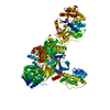 6mfyC 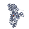 6mfzC S: Starting model for refinement C: citing same article ( |
|---|---|
| Similar structure data |

-Links
 PDBj
PDBj


- Assembly
| Deposited unit | 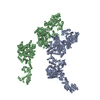
| ||||||||||
|---|---|---|---|---|---|---|---|---|---|---|---|
| 1 |
| ||||||||||
| 2 |
| ||||||||||
| Unit cell |
|
-Components
| #1: Protein | Mass: 196107.047 Da / Num. of mol.: 2 / Fragment: UNP residues 2-1716 Source method: isolated from a genetically manipulated source Source: (gene. exp.) Brevibacillus parabrevis (bacteria) / Gene: lgrA / Production host: #2: Chemical |   Mass: 785.827 Da / Num. of mol.: 2 / Source method: obtained synthetically / Formula: C27H48N9O12PS2 Mass: 785.827 Da / Num. of mol.: 2 / Source method: obtained synthetically / Formula: C27H48N9O12PS2Has protein modification | Y | Nonpolymer details | DG9 is deprotonated. | |
|---|
-Experimental details
-Experiment
| Experiment | Method: X-RAY DIFFRACTION / Number of used crystals: 1 |
|---|
- Sample preparation
| Crystal | Density Matthews: 5.78 Å3/Da / Density % sol: 78.73 % |
|---|---|
| Crystal grow | Temperature: 277 K / Method: vapor diffusion, sitting drop / pH: 6.8 Details: 0.25 M sodium fluoride, 3.1 M sodium formate, pH 6.8 |
-Data collection
| Diffraction | Mean temperature: 100 K / Serial crystal experiment: N |
|---|---|
| Diffraction source | Source: SYNCHROTRON / Site: APS  / Beamline: 24-ID-C / Wavelength: 0.979 Å / Beamline: 24-ID-C / Wavelength: 0.979 Å |
| Detector | Type: DECTRIS PILATUS 6M-F / Detector: PIXEL / Date: Mar 12, 2017 |
| Radiation | Monochromator: cryo-cooled double crystal Si(111) / Protocol: SINGLE WAVELENGTH / Monochromatic (M) / Laue (L): M / Scattering type: x-ray |
| Radiation wavelength | Wavelength: 0.979 Å / Relative weight: 1 |
| Reflection | Resolution: 6→78.44 Å / Num. obs: 22451 / % possible obs: 99.8 % / Redundancy: 6.3 % / Biso Wilson estimate: 280.3 Å2 / Net I/σ(I): 6.9 |
| Reflection shell | Resolution: 6→6.48 Å / Mean I/σ(I) obs: 1.6 / Num. unique obs: 4552 / CC1/2: 0.451 |
- Processing
| Software |
| |||||||||||||||||||||||||||||||||||||||||||||||||||||||||||||||
|---|---|---|---|---|---|---|---|---|---|---|---|---|---|---|---|---|---|---|---|---|---|---|---|---|---|---|---|---|---|---|---|---|---|---|---|---|---|---|---|---|---|---|---|---|---|---|---|---|---|---|---|---|---|---|---|---|---|---|---|---|---|---|---|---|
| Refinement | Method to determine structure: MOLECULAR REPLACEMENT Starting model: PDB entry 6MFW Resolution: 6→78.44 Å / SU ML: 0.95 / Cross valid method: FREE R-VALUE / σ(F): 1.36 / Phase error: 32.65
| |||||||||||||||||||||||||||||||||||||||||||||||||||||||||||||||
| Solvent computation | Shrinkage radii: 0.9 Å / VDW probe radii: 1.11 Å | |||||||||||||||||||||||||||||||||||||||||||||||||||||||||||||||
| Refinement step | Cycle: LAST / Resolution: 6→78.44 Å
| |||||||||||||||||||||||||||||||||||||||||||||||||||||||||||||||
| Refine LS restraints |
| |||||||||||||||||||||||||||||||||||||||||||||||||||||||||||||||
| LS refinement shell |
|
