 Movie
Movie Controller

[English] 日本語
 Yorodumi
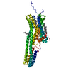
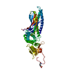
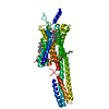
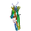
- PDB-6me9: XFEL crystal structure of human melatonin receptor MT2 in complex... -
+ Open data

- Basic information
| Entry | Database: PDB / ID: 6me9 | ||||||
|---|---|---|---|---|---|---|---|
| Title | XFEL crystal structure of human melatonin receptor MT2 in complex with ramelteon | ||||||
 Components Components | Soluble cytochrome b562,Melatonin receptor type 1B,Rubredoxin | ||||||
 Keywords Keywords | MEMBRANE PROTEIN / GPCR / melatonin receptor type 1B (MT2) / ramelteon / XFEL / LCP / BRIL / Rubredoxin / circadian rhythm / jetlag / type 2 diabetes | ||||||
| Function / homology |  Function and homology information Function and homology informationmelatonin receptor activity / positive regulation of circadian sleep/wake cycle, non-REM sleep / positive regulation of transmission of nerve impulse / negative regulation of transmission of nerve impulse / regulation of neuronal action potential / alkane catabolic process / camera-type eye development / Class A/1 (Rhodopsin-like receptors) / negative regulation of receptor guanylyl cyclase signaling pathway / negative regulation of vasoconstriction ...melatonin receptor activity / positive regulation of circadian sleep/wake cycle, non-REM sleep / positive regulation of transmission of nerve impulse / negative regulation of transmission of nerve impulse / regulation of neuronal action potential / alkane catabolic process / camera-type eye development / Class A/1 (Rhodopsin-like receptors) / negative regulation of receptor guanylyl cyclase signaling pathway / negative regulation of vasoconstriction / negative regulation of cytosolic calcium ion concentration / regulation of insulin secretion / negative regulation of insulin secretion / G protein-coupled receptor signaling pathway, coupled to cyclic nucleotide second messenger / electron transport chain / G protein-coupled receptor activity / glucose homeostasis / chemical synaptic transmission / negative regulation of neuron apoptotic process / G alpha (i) signalling events / periplasmic space / electron transfer activity / iron ion binding / G protein-coupled receptor signaling pathway / heme binding / synapse / plasma membrane Similarity search - Function | ||||||
| Biological species |   Homo sapiens (human)Clostridium pasteurianum (bacteria) Homo sapiens (human)Clostridium pasteurianum (bacteria) | ||||||
| Method |  X-RAY DIFFRACTION / FREE ELECTRON LASER / MOLECULAR REPLACEMENT / Resolution: 3.3 Å X-RAY DIFFRACTION / FREE ELECTRON LASER / MOLECULAR REPLACEMENT / Resolution: 3.3 Å | ||||||
 Authors Authors | Johansson, L.C. / Stauch, B. / McCorvy, J. / Han, G.W. / Patel, N. / Batyuk, A. / Gati, C. / Li, C. / Grandner, J. / Hao, S. ...Johansson, L.C. / Stauch, B. / McCorvy, J. / Han, G.W. / Patel, N. / Batyuk, A. / Gati, C. / Li, C. / Grandner, J. / Hao, S. / Olsen, R.H.J. / Tribo, A.R. / Zaare, S. / Zhu, L. / Zatsepin, N.A. / Weierstall, U. / Liu, W. / Roth, B.L. / Katritch, V. / Cherezov, V. | ||||||
| Funding support |  United States, 1items United States, 1items
| ||||||
 Citation Citation | Journal: Nature / Year: 2019 Title: XFEL structures of the human MT2melatonin receptor reveal the basis of subtype selectivity. Authors: Johansson, L.C. / Stauch, B. / McCorvy, J.D. / Han, G.W. / Patel, N. / Huang, X.P. / Batyuk, A. / Gati, C. / Slocum, S.T. / Li, C. / Grandner, J.M. / Hao, S. / Olsen, R.H.J. / Tribo, A.R. / ...Authors: Johansson, L.C. / Stauch, B. / McCorvy, J.D. / Han, G.W. / Patel, N. / Huang, X.P. / Batyuk, A. / Gati, C. / Slocum, S.T. / Li, C. / Grandner, J.M. / Hao, S. / Olsen, R.H.J. / Tribo, A.R. / Zaare, S. / Zhu, L. / Zatsepin, N.A. / Weierstall, U. / Yous, S. / Stevens, R.C. / Liu, W. / Roth, B.L. / Katritch, V. / Cherezov, V. | ||||||
| History |
|
- Structure visualization
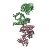
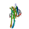
| Structure viewer | Molecule: MolmilJmol/JSmol |
|---|
- Downloads & links
-Download
| PDBx/mmCIF format | 6me9.cif.gz | 170.5 KB | Display | PDBx/mmCIF format |
|---|---|---|---|---|
| PDB format | pdb6me9.ent.gz | 128.5 KB | Display | PDB format |
| PDBx/mmJSON format | 6me9.json.gz | Tree view | PDBx/mmJSON format | |
| Others |  Other downloads Other downloads |
-Validation report
| Arichive directory | https://data.pdbj.org/pub/pdb/validation_reports/me/6me9ftp://data.pdbj.org/pub/pdb/validation_reports/me/6me9 | HTTPS FTP |
|---|
-Related structure data
| Related structure data |  6me6C  6me7C  6me8C  1iroS S: Starting model for refinement C: citing same article ( |
|---|---|
| Similar structure data |


-Links
 PDBj
PDBj


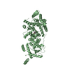
- Assembly
| Deposited unit | 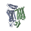
| ||||||||
|---|---|---|---|---|---|---|---|---|---|
| 1 | 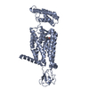
| ||||||||
| 2 | 
| ||||||||
| Unit cell |
| ||||||||
| Details | BIOLOGICAL UNIT IS UNKNOWN |
-Components
| #1: Protein | Mass: 51514.578 Da / Num. of mol.: 2 Mutation: M2007W, H2102I, R2106L, P37S, D86N, L108F, F129W, N137D, C140L, W246F, A305P Source method: isolated from a genetically manipulated source Source: (gene. exp.) Homo sapiens (human), (gene. exp.) Clostridium pasteurianum (bacteria)Gene: cybC, MTNR1B / Production host:   Spodoptera frugiperda (fall armyworm) Spodoptera frugiperda (fall armyworm)References: UniProt: P0ABE7, UniProt: P49286, UniProt: P00268 #2: Chemical | 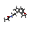  Mass: 259.343 Da / Num. of mol.: 2 / Source method: obtained synthetically / Formula: C16H21NO2 / Comment: medication, agonist*YM Mass: 259.343 Da / Num. of mol.: 2 / Source method: obtained synthetically / Formula: C16H21NO2 / Comment: medication, agonist*YM#3: Chemical | ChemComp-ZN / |   Mass: 65.409 Da / Num. of mol.: 1 / Source method: obtained synthetically / Formula: Zn Mass: 65.409 Da / Num. of mol.: 1 / Source method: obtained synthetically / Formula: ZnHas protein modification | Y | |
|---|
-Experimental details
-Experiment
| Experiment | Method: X-RAY DIFFRACTION / Number of used crystals: 1 |
|---|
- Sample preparation
| Crystal | Density Matthews: 3.64 Å3/Da / Density % sol: 66.19 % |
|---|---|
| Crystal grow | Temperature: 293 K / Method: lipidic cubic phase Details: N-(2-Acetamido)iminodiacetic acid, PEG 400, ammonium acetate |
-Data collection
| Diffraction | Mean temperature: 293 K / Serial crystal experiment: Y |
|---|---|
| Diffraction source | Source: FREE ELECTRON LASER / Site: SLAC LCLS / Beamline: CXI / Wavelength: 1.3 Å |
| Detector | Type: CS-PAD CXI-1 / Detector: PIXEL / Date: Dec 7, 2017 |
| Radiation | Protocol: SINGLE WAVELENGTH / Monochromatic (M) / Laue (L): M / Scattering type: x-ray |
| Radiation wavelength | Wavelength: 1.3 Å / Relative weight: 1 |
| Reflection | Resolution: 3.3→22 Å / Num. obs: 22143 / % possible obs: 100 % / Redundancy: 221.1 % / Biso Wilson estimate: 79.55 Å2 / Net I/σ(I): 3.67 |
| Reflection shell | Resolution: 3.3→3.46 Å / Redundancy: 84.6 % / Num. unique obs: 2938 / R split: 0.2 / % possible all: 100 |
| Serial crystallography measurement | Pulse duration: 30 fsec. / Pulse energy: 2.1 µJ / Pulse photon energy: 9.83 keV / XFEL pulse repetition rate: 120 Hz |
| Serial crystallography sample delivery | Method: injection |
| Serial crystallography sample delivery injection | Carrier solvent: LCP / Flow rate: 0.357 µL/min / Injector diameter: 50 µm / Injector nozzle: LCP / Power by: HPLC pump |
| Serial crystallography data reduction | Crystal hits: 60005 / Frames indexed: 28834 / Frames total: 727004 |
- Processing
| Software |
| ||||||||||||||||||||||||||||||||||||||||||||||||||||||||||||||||||||||||||||||||||||||||||||||||||||||||||||||||||
|---|---|---|---|---|---|---|---|---|---|---|---|---|---|---|---|---|---|---|---|---|---|---|---|---|---|---|---|---|---|---|---|---|---|---|---|---|---|---|---|---|---|---|---|---|---|---|---|---|---|---|---|---|---|---|---|---|---|---|---|---|---|---|---|---|---|---|---|---|---|---|---|---|---|---|---|---|---|---|---|---|---|---|---|---|---|---|---|---|---|---|---|---|---|---|---|---|---|---|---|---|---|---|---|---|---|---|---|---|---|---|---|---|---|---|---|
| Refinement | Method to determine structure: MOLECULAR REPLACEMENT Starting model: PDB id:1IRO Resolution: 3.3→22 Å / Cor.coef. Fo:Fc: 0.882 / Cor.coef. Fo:Fc free: 0.87 / Rfactor Rfree error: 0 / Cross valid method: THROUGHOUT / σ(F): 0 / SU Rfree Blow DPI: 0.441
| ||||||||||||||||||||||||||||||||||||||||||||||||||||||||||||||||||||||||||||||||||||||||||||||||||||||||||||||||||
| Displacement parameters | Biso mean: 136.26 Å2
| ||||||||||||||||||||||||||||||||||||||||||||||||||||||||||||||||||||||||||||||||||||||||||||||||||||||||||||||||||
| Refine analyze | Luzzati coordinate error obs: 0.83 Å | ||||||||||||||||||||||||||||||||||||||||||||||||||||||||||||||||||||||||||||||||||||||||||||||||||||||||||||||||||
| Refinement step | Cycle: 1 / Resolution: 3.3→22 Å
| ||||||||||||||||||||||||||||||||||||||||||||||||||||||||||||||||||||||||||||||||||||||||||||||||||||||||||||||||||
| Refine LS restraints |
| ||||||||||||||||||||||||||||||||||||||||||||||||||||||||||||||||||||||||||||||||||||||||||||||||||||||||||||||||||
| LS refinement shell | Resolution: 3.3→3.46 Å / Rfactor Rfree error: 0 / Total num. of bins used: 11
|
