 Movie
Movie Controller

[English] 日本語
 Yorodumi
- PDB-6mdf: Mevalonate kinase from Methanosarcina mazei with 5-phosphomevalon... -
+ Open data

- Basic information
| Entry | Database: PDB / ID: 6mdf | ||||||
|---|---|---|---|---|---|---|---|
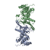
| Title | Mevalonate kinase from Methanosarcina mazei with 5-phosphomevalonate bound | ||||||
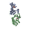
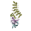
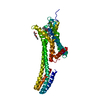
 Components Components | Mevalonate kinase | ||||||
 Keywords Keywords | TRANSFERASE / kinase | ||||||
| Function / homology |  Function and homology information Function and homology informationmevalonate kinase / mevalonate kinase activity / isopentenyl diphosphate biosynthetic process, mevalonate pathway / magnesium ion binding / ATP binding / cytosol Similarity search - Function | ||||||
| Biological species |  Methanosarcina mazei (archaea) Methanosarcina mazei (archaea) | ||||||
| Method |  X-RAY DIFFRACTION / SYNCHROTRON / MOLECULAR REPLACEMENT / Resolution: 2.46 Å X-RAY DIFFRACTION / SYNCHROTRON / MOLECULAR REPLACEMENT / Resolution: 2.46 Å | ||||||
 Authors Authors | Miller, B.R. / Kung, Y. | ||||||
| Funding support |  United States, 1items United States, 1items
| ||||||
 Citation Citation | Journal: PLoS ONE / Year: 2018 Title: Structural insight into substrate and product binding in an archaeal mevalonate kinase. Authors: Miller, B.R. / Kung, Y. | ||||||
| History |
|
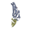
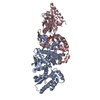
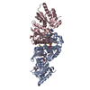
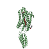
- Structure visualization
| Structure viewer | Molecule: MolmilJmol/JSmol |
|---|
- Downloads & links
-Download
| PDBx/mmCIF format | 6mdf.cif.gz | 129.1 KB | Display | PDBx/mmCIF format |
|---|---|---|---|---|
| PDB format | pdb6mdf.ent.gz | 98.3 KB | Display | PDB format |
| PDBx/mmJSON format | 6mdf.json.gz | Tree view | PDBx/mmJSON format | |
| Others |  Other downloads Other downloads |
-Validation report
| Arichive directory | https://data.pdbj.org/pub/pdb/validation_reports/md/6mdfftp://data.pdbj.org/pub/pdb/validation_reports/md/6mdf | HTTPS FTP |
|---|
-Related structure data
| Related structure data | 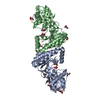 6mdeC 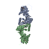 4hacS S: Starting model for refinement C: citing same article ( |
|---|---|
| Similar structure data |

-Links
 PDBj
PDBj- Assembly
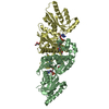
| Deposited unit | 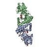
| ||||||||
|---|---|---|---|---|---|---|---|---|---|
| 1 |
| ||||||||
| Unit cell |
|
-Components
| #1: Protein | Mass: 31637.281 Da / Num. of mol.: 2 Source method: isolated from a genetically manipulated source Source: (gene. exp.) Methanosarcina mazei (archaea) / Gene: mvk, MM_1762 / Production host:  #2: Chemical | 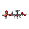  Mass: 228.137 Da / Num. of mol.: 2 / Source method: obtained synthetically / Formula: C6H13O7P / Feature type: SUBJECT OF INVESTIGATION Mass: 228.137 Da / Num. of mol.: 2 / Source method: obtained synthetically / Formula: C6H13O7P / Feature type: SUBJECT OF INVESTIGATION#3: Chemical | ChemComp-GOL /   Mass: 92.094 Da / Num. of mol.: 13 / Source method: obtained synthetically / Formula: C3H8O3 Mass: 92.094 Da / Num. of mol.: 13 / Source method: obtained synthetically / Formula: C3H8O3#4: Water | ChemComp-HOH / |  Mass: 18.015 Da / Num. of mol.: 208 / Source method: isolated from a natural source / Formula: H2O Mass: 18.015 Da / Num. of mol.: 208 / Source method: isolated from a natural source / Formula: H2O |
|---|
-Experimental details
-Experiment
| Experiment | Method: X-RAY DIFFRACTION / Number of used crystals: 1 |
|---|
- Sample preparation
| Crystal | Density Matthews: 2.37 Å3/Da / Density % sol: 48.18 % |
|---|---|
| Crystal grow | Temperature: 298 K / Method: vapor diffusion, hanging drop / pH: 5.5 Details: 100 mM Bis-Tris, pH 5.5, 200-300 mM sodium potassium tartrate, 15-25% PEG8000 |
-Data collection
| Diffraction | Mean temperature: 100 K |
|---|---|
| Diffraction source | Source: SYNCHROTRON / Site: APS / Beamline: 24-ID-E / Wavelength: 0.9792 Å |
| Detector | Type: DECTRIS EIGER X 16M / Detector: PIXEL / Date: Mar 30, 2018 |
| Radiation | Monochromator: Cryogenically-cooled single crystal Si(220) side bounce Protocol: SINGLE WAVELENGTH / Monochromatic (M) / Laue (L): M / Scattering type: x-ray |
| Radiation wavelength | Wavelength: 0.9792 Å / Relative weight: 1 |
| Reflection | Resolution: 2.46→51.92 Å / Num. obs: 22260 / % possible obs: 96.95 % / Redundancy: 4.3 % / CC1/2: 0.999 / Rmerge(I) obs: 0.1031 / Net I/σ(I): 8.73 |
| Reflection shell | Resolution: 2.46→2.55 Å / Redundancy: 4 % / Rmerge(I) obs: 0.3752 / Mean I/σ(I) obs: 2.85 / Num. unique obs: 2135 / CC1/2: 0.843 / % possible all: 96.3 |
- Processing
| Software |
| |||||||||||||||||||||||||||||||||||||||||||||||||||||||||||||||
|---|---|---|---|---|---|---|---|---|---|---|---|---|---|---|---|---|---|---|---|---|---|---|---|---|---|---|---|---|---|---|---|---|---|---|---|---|---|---|---|---|---|---|---|---|---|---|---|---|---|---|---|---|---|---|---|---|---|---|---|---|---|---|---|---|
| Refinement | Method to determine structure: MOLECULAR REPLACEMENT Starting model: PDB entry 4HAC Resolution: 2.46→51.92 Å / SU ML: 0.35 / Cross valid method: FREE R-VALUE / σ(F): 1.34 / Phase error: 26.84
| |||||||||||||||||||||||||||||||||||||||||||||||||||||||||||||||
| Solvent computation | Shrinkage radii: 0.9 Å / VDW probe radii: 1.11 Å | |||||||||||||||||||||||||||||||||||||||||||||||||||||||||||||||
| Refinement step | Cycle: LAST / Resolution: 2.46→51.92 Å
| |||||||||||||||||||||||||||||||||||||||||||||||||||||||||||||||
| Refine LS restraints |
| |||||||||||||||||||||||||||||||||||||||||||||||||||||||||||||||
| LS refinement shell |
|
