 Movie
Movie Controller


+ Open data

- Basic information
| Entry | Database: PDB / ID: 6mb2 | ||||||||||||||||||||||||
|---|---|---|---|---|---|---|---|---|---|---|---|---|---|---|---|---|---|---|---|---|---|---|---|---|---|
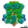
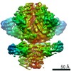
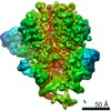
| Title | Cryo-EM structure of the PYD filament of AIM2 | ||||||||||||||||||||||||
 Components Components |
| ||||||||||||||||||||||||
 Keywords Keywords | IMMUNE SYSTEM / Filament / higher order / innate immunity / inflammasome | ||||||||||||||||||||||||
| Function / homology |  Function and homology information Function and homology informationpyroptosome complex assembly / AIM2 inflammasome complex assembly / The AIM2 inflammasome / AIM2 inflammasome complex / regulation of behavior / cysteine-type endopeptidase activator activity / Cytosolic sensors of pathogen-associated DNA / : / pattern recognition receptor signaling pathway / pattern recognition receptor activity ...pyroptosome complex assembly / AIM2 inflammasome complex assembly / The AIM2 inflammasome / AIM2 inflammasome complex / regulation of behavior / cysteine-type endopeptidase activator activity / Cytosolic sensors of pathogen-associated DNA / : / pattern recognition receptor signaling pathway / pattern recognition receptor activity / T cell homeostasis / pyroptotic inflammatory response / cellular response to interferon-beta / positive regulation of defense response to virus by host / activation of innate immune response / signaling adaptor activity / negative regulation of phosphatidylinositol 3-kinase/protein kinase B signal transduction / bioluminescence / : / tumor necrosis factor-mediated signaling pathway / positive regulation of interleukin-1 beta production / generation of precursor metabolites and energy / protein maturation / neuron cellular homeostasis / brain development / cellular response to xenobiotic stimulus / positive regulation of inflammatory response / site of double-strand break / double-stranded DNA binding / defense response to virus / immune response / innate immune response / DNA damage response / mitochondrion / nucleoplasm / identical protein binding / cytoplasm / cytosol Similarity search - Function | ||||||||||||||||||||||||
| Biological species |  Homo sapiens (human) Homo sapiens (human)  Aequorea victoria (jellyfish) Aequorea victoria (jellyfish) | ||||||||||||||||||||||||
| Method | ELECTRON MICROSCOPY / helical reconstruction / cryo EM / Resolution: 5 Å | ||||||||||||||||||||||||
 Authors Authors | Lu, A. / Li, Y. / Wu, H. | ||||||||||||||||||||||||
 Citation Citation | Journal: Cell Discov / Year: 2015 Title: Plasticity in PYD assembly revealed by cryo-EM structure of the PYD filament of AIM2. Authors: Alvin Lu / Yang Li / Qian Yin / Jianbin Ruan / Xiong Yu / Edward Egelman / Hao Wu /  Abstract: Absent in melanoma 2 (AIM2) is an essential cytosolic double-stranded DNA receptor that assembles with the adaptor, apoptosis-associated speck-like protein containing a caspase recruitment domain ...Absent in melanoma 2 (AIM2) is an essential cytosolic double-stranded DNA receptor that assembles with the adaptor, apoptosis-associated speck-like protein containing a caspase recruitment domain (ASC), and caspase-1 to form the AIM2 inflammasome, which leads to proteolytic maturation of cytokines and pyroptotic cell death. AIM2 contains an N-terminal Pyrin domain (PYD) that interacts with ASC through PYD/PYD interactions and nucleates ASC filament formation. To elucidate the molecular basis of AIM2-induced ASC polymerization, we generated AIM2 filaments fused to green fluorescent protein (GFP) and determined its cryo-electron microscopic (cryo-EM) structure. The map showed distinct definition of helices, allowing fitting of the crystal structure. Surprisingly, the GFP-AIM2 filament is a 1-start helix with helical parameters distinct from those of the 3-start ASC filament. However, despite the apparent symmetry difference, helical net and detailed interface analyses reveal minimal changes in subunit packing. GFP-AIM2 nucleated ASC filament formation in comparable efficiency as untagged AIM2, suggesting assembly plasticity in both AIM2 and ASC. The DNA-binding domain of AIM2 is able to form AIM2/DNA filaments, within which the AIM2 is brought into proximity to template ASC filament assembly. Because ASC is able to interact with many PYD-containing receptors for the formation of inflammasomes, the observed structural plasticity may be critically important for this versatility in the PYD/PYD interactions. | ||||||||||||||||||||||||
| History |
|
- Structure visualization
| Movie |
Movie viewer |
|---|---|
| Structure viewer | Molecule: MolmilJmol/JSmol |
- Downloads & links
-Download
| PDBx/mmCIF format | 6mb2.cif.gz | 916.2 KB | Display | PDBx/mmCIF format |
|---|---|---|---|---|
| PDB format | pdb6mb2.ent.gz | 759.9 KB | Display | PDB format |
| PDBx/mmJSON format | 6mb2.json.gz | Tree view | PDBx/mmJSON format | |
| Others |  Other downloads Other downloads |
-Validation report
| Arichive directory | https://data.pdbj.org/pub/pdb/validation_reports/mb/6mb2ftp://data.pdbj.org/pub/pdb/validation_reports/mb/6mb2 | HTTPS FTP |
|---|
-Related structure data
| Related structure data | 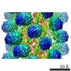 9064MC M: map data used to model this data C: citing same article ( |
|---|---|
| Similar structure data |


-Links
 PDBj
PDBj


- Assembly
| Deposited unit | 
|
|---|---|
| 1 |
|
| Symmetry | Helical symmetry: (Circular symmetry: 1 / Dyad axis: no / N subunits divisor: 1 / Num. of operations: 15 / Rise per n subunits: 6 Å / Rotation per n subunits: 138.9 °) |
-Components
| #1: Protein | Mass: 10839.629 Da / Num. of mol.: 15 / Fragment: PYD (UNP residues 1-93) Source method: isolated from a genetically manipulated source Source: (gene. exp.) Homo sapiens (human) / Gene: AIM2 / Production host:  #2: Protein | Mass: 25924.553 Da / Num. of mol.: 15 / Fragment: UNP residues 2-229 Source method: isolated from a genetically manipulated source Source: (gene. exp.) Aequorea victoria (jellyfish) / Gene: GFP / Production host: Has protein modification | Y | |
|---|
-Experimental details
-Experiment
| Experiment | Method: ELECTRON MICROSCOPY |
|---|---|
| EM experiment | Aggregation state: FILAMENT / 3D reconstruction method: helical reconstruction |
- Sample preparation
| Component | Name: PYD of AIM2 / Type: ORGANELLE OR CELLULAR COMPONENT / Entity ID: all / Source: RECOMBINANT |
|---|---|
| Source (natural) | Organism: Homo sapiens (human) |
| Source (recombinant) | Organism: |
| Buffer solution | pH: 8 |
| Specimen | Embedding applied: NO / Shadowing applied: NO / Staining applied: NO / Vitrification applied: YES |
| Specimen support | Details: unspecified |
| Vitrification | Cryogen name: ETHANE |
- Electron microscopy imaging
| Experimental equipment |  Model: Titan Krios / Image courtesy: FEI Company |
|---|---|
| Microscopy | Model: FEI TITAN KRIOS |
| Electron gun | Electron source:  FIELD EMISSION GUN / Accelerating voltage: 300 kV / Illumination mode: FLOOD BEAM FIELD EMISSION GUN / Accelerating voltage: 300 kV / Illumination mode: FLOOD BEAM |
| Electron lens | Mode: BRIGHT FIELD |
| Image recording | Electron dose: 50 e/Å2 / Film or detector model: FEI FALCON II (4k x 4k) |
- Processing
| Software | Name: PHENIX / Version: 1.8.2_1309 / Classification: refinement | ||||||||||||||||||||||||||||||||||||||||||||||||||||||||||||||||||||||||||||||||||||||||||||||||||||||||||||||||||||||||||||||||||||||||||||
|---|---|---|---|---|---|---|---|---|---|---|---|---|---|---|---|---|---|---|---|---|---|---|---|---|---|---|---|---|---|---|---|---|---|---|---|---|---|---|---|---|---|---|---|---|---|---|---|---|---|---|---|---|---|---|---|---|---|---|---|---|---|---|---|---|---|---|---|---|---|---|---|---|---|---|---|---|---|---|---|---|---|---|---|---|---|---|---|---|---|---|---|---|---|---|---|---|---|---|---|---|---|---|---|---|---|---|---|---|---|---|---|---|---|---|---|---|---|---|---|---|---|---|---|---|---|---|---|---|---|---|---|---|---|---|---|---|---|---|---|---|---|
| CTF correction | Type: PHASE FLIPPING AND AMPLITUDE CORRECTION | ||||||||||||||||||||||||||||||||||||||||||||||||||||||||||||||||||||||||||||||||||||||||||||||||||||||||||||||||||||||||||||||||||||||||||||
| Helical symmerty | Angular rotation/subunit: 138.9 ° / Axial rise/subunit: 6 Å / Axial symmetry: C1 | ||||||||||||||||||||||||||||||||||||||||||||||||||||||||||||||||||||||||||||||||||||||||||||||||||||||||||||||||||||||||||||||||||||||||||||
| 3D reconstruction | Resolution: 5 Å / Resolution method: FSC 0.143 CUT-OFF / Num. of particles: 54973 / Symmetry type: HELICAL | ||||||||||||||||||||||||||||||||||||||||||||||||||||||||||||||||||||||||||||||||||||||||||||||||||||||||||||||||||||||||||||||||||||||||||||
| Refinement | Resolution: 5→150 Å / SU ML: 1.77 / σ(F): 0.76 / Phase error: 64.53 / Stereochemistry target values: MLHL
| ||||||||||||||||||||||||||||||||||||||||||||||||||||||||||||||||||||||||||||||||||||||||||||||||||||||||||||||||||||||||||||||||||||||||||||
| Solvent computation | Shrinkage radii: 0.9 Å / VDW probe radii: 1.11 Å / Solvent model: FLAT BULK SOLVENT MODEL | ||||||||||||||||||||||||||||||||||||||||||||||||||||||||||||||||||||||||||||||||||||||||||||||||||||||||||||||||||||||||||||||||||||||||||||
| Refine LS restraints |
| ||||||||||||||||||||||||||||||||||||||||||||||||||||||||||||||||||||||||||||||||||||||||||||||||||||||||||||||||||||||||||||||||||||||||||||
| LS refinement shell |
|
