 Movie
Movie Controller

[English] 日本語
 Yorodumi
- PDB-6mao: Crystal structure of Deoxyuridine 5'-triphosphate nucleotidohydro... -
+ Open data

- Basic information
| Entry | Database: PDB / ID: 6mao | ||||||
|---|---|---|---|---|---|---|---|
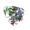
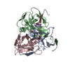
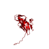
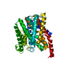
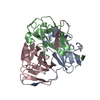
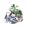
| Title | Crystal structure of Deoxyuridine 5'-triphosphate nucleotidohydrolase from Legionella pneumophila Philadelphia 1 in complex with dUMP (Deoxyuridine 5'-monophosphate) | ||||||
 Components Components | Deoxyuridine 5'-triphosphate nucleotidohydrolase | ||||||
 Keywords Keywords | HYDROLASE / SSGCID / Structural Genomics / Seattle Structural Genomics Center for Infectious Disease | ||||||
| Function / homology |  Function and homology information Function and homology informationdUTP catabolic process / dUMP biosynthetic process / dUTP diphosphatase / dUTP diphosphatase activity / magnesium ion binding Similarity search - Function | ||||||
| Biological species |  Legionella pneumophila subsp. pneumophila (bacteria) Legionella pneumophila subsp. pneumophila (bacteria) | ||||||
| Method |  X-RAY DIFFRACTION / MOLECULAR REPLACEMENT / Resolution: 1.95 Å X-RAY DIFFRACTION / MOLECULAR REPLACEMENT / Resolution: 1.95 Å | ||||||
 Authors Authors | Seattle Structural Genomics Center for Infectious Disease (SSGCID) | ||||||
 Citation Citation | Journal: Acta Crystallogr.,Sect.F / Year: 2025 Title: Structural characterization of dUTPase from Legionella pneumophila Authors: Nguyen, C.L. / Tramell, A.R. / Norman, J.O. / Abendroth, J. / Barrett, K.F. / Craig, J.K. / Edwards, T.E. / Lorimer, D.D. / McLaughlin, K.J. | ||||||
| History |
|
- Structure visualization
| Structure viewer | Molecule: MolmilJmol/JSmol |
|---|
- Downloads & links
-Download
| PDBx/mmCIF format | 6mao.cif.gz | 75.6 KB | Display | PDBx/mmCIF format |
|---|---|---|---|---|
| PDB format | pdb6mao.ent.gz | 54.4 KB | Display | PDB format |
| PDBx/mmJSON format | 6mao.json.gz | Tree view | PDBx/mmJSON format | |
| Others |  Other downloads Other downloads |
-Validation report
| Arichive directory | https://data.pdbj.org/pub/pdb/validation_reports/ma/6maoftp://data.pdbj.org/pub/pdb/validation_reports/ma/6mao | HTTPS FTP |
|---|
-Related structure data
| Related structure data |  6maiSC S: Starting model for refinement C: citing same article ( |
|---|---|
| Similar structure data | |
| Other databases |

-Links
 PDBj
PDBj
- Assembly
| Deposited unit | 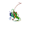
| ||||||||
|---|---|---|---|---|---|---|---|---|---|
| 1 | 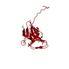
| ||||||||
| Unit cell |
| ||||||||
| Components on special symmetry positions |
|
-Components
| #1: Protein | Mass: 17391.789 Da / Num. of mol.: 1 Source method: isolated from a genetically manipulated source Source: (gene. exp.) Legionella pneumophila subsp. pneumophila (strain Philadelphia 1 / ATCC 33152 / DSM 7513) (bacteria)Strain: Philadelphia 1 / ATCC 33152 / DSM 7513 / Gene: dut, lpg2487 / Plasmid: LepnA.01206.a.B1 Production host: References: UniProt: Q5ZSN0, dUTP diphosphatase |
|---|---|
| #2: Chemical | ChemComp-UMP /   Mass: 308.182 Da / Num. of mol.: 1 / Source method: obtained synthetically / Formula: C9H13N2O8P Mass: 308.182 Da / Num. of mol.: 1 / Source method: obtained synthetically / Formula: C9H13N2O8P |
| #3: Chemical | ChemComp-MPD / (  Mass: 118.174 Da / Num. of mol.: 1 / Source method: obtained synthetically / Formula: C6H14O2 / Comment: precipitant*YM Mass: 118.174 Da / Num. of mol.: 1 / Source method: obtained synthetically / Formula: C6H14O2 / Comment: precipitant*YM |
| #4: Chemical | ChemComp-SO4 /   Mass: 96.063 Da / Num. of mol.: 1 / Source method: obtained synthetically / Formula: SO4 Mass: 96.063 Da / Num. of mol.: 1 / Source method: obtained synthetically / Formula: SO4 |
| #5: Water | ChemComp-HOH /  Mass: 18.015 Da / Num. of mol.: 134 / Source method: isolated from a natural source / Formula: H2O Mass: 18.015 Da / Num. of mol.: 134 / Source method: isolated from a natural source / Formula: H2O |
| Has protein modification | N |
-Experimental details
-Experiment
| Experiment | Method: X-RAY DIFFRACTION / Number of used crystals: 1 |
|---|
- Sample preparation
| Crystal | Density Matthews: 3.38 Å3/Da / Density % sol: 63.6 % |
|---|---|
| Crystal grow | Temperature: 285 K / Method: vapor diffusion, sitting drop / pH: 6.5 Details: Molecular Dimensions Morpheus screen, condition C4: 12.5% w/v PEG 1000, 12.5% w/v PEG 3350, 12.5% v/v MPD 30mM each sodium nitrate, disodium hydrogen phosphate, ammonium sulfate: 100mM ...Details: Molecular Dimensions Morpheus screen, condition C4: 12.5% w/v PEG 1000, 12.5% w/v PEG 3350, 12.5% v/v MPD 30mM each sodium nitrate, disodium hydrogen phosphate, ammonium sulfate: 100mM MES/imidazole pH 6.5: LepnA.01206.a.B1.PS38438 at 24.33mg/ml: 2h soak with 5mM MgCl2 + dUTP with was converted to dUMP: cryo: direct: EG in two steps: tray 301633a1, puck PXP9-8 |
-Data collection
| Diffraction | Mean temperature: 100 K | ||||||||||||||||||||||||||||||||||||||||||||||||||||||||||||||||||||||||||||||||||||||||||||||||||||||||||||||||||||||||||||||||||||||||||||||||||||||||||||||||||||||||
|---|---|---|---|---|---|---|---|---|---|---|---|---|---|---|---|---|---|---|---|---|---|---|---|---|---|---|---|---|---|---|---|---|---|---|---|---|---|---|---|---|---|---|---|---|---|---|---|---|---|---|---|---|---|---|---|---|---|---|---|---|---|---|---|---|---|---|---|---|---|---|---|---|---|---|---|---|---|---|---|---|---|---|---|---|---|---|---|---|---|---|---|---|---|---|---|---|---|---|---|---|---|---|---|---|---|---|---|---|---|---|---|---|---|---|---|---|---|---|---|---|---|---|---|---|---|---|---|---|---|---|---|---|---|---|---|---|---|---|---|---|---|---|---|---|---|---|---|---|---|---|---|---|---|---|---|---|---|---|---|---|---|---|---|---|---|---|---|---|---|
| Diffraction source | Source: ROTATING ANODE / Type: RIGAKU FR-E+ SUPERBRIGHT / Wavelength: 1.5418 Å | ||||||||||||||||||||||||||||||||||||||||||||||||||||||||||||||||||||||||||||||||||||||||||||||||||||||||||||||||||||||||||||||||||||||||||||||||||||||||||||||||||||||||
| Detector | Type: RIGAKU SATURN 944+ / Detector: CCD / Date: Aug 21, 2018 | ||||||||||||||||||||||||||||||||||||||||||||||||||||||||||||||||||||||||||||||||||||||||||||||||||||||||||||||||||||||||||||||||||||||||||||||||||||||||||||||||||||||||
| Radiation | Protocol: SINGLE WAVELENGTH / Monochromatic (M) / Laue (L): M / Scattering type: x-ray | ||||||||||||||||||||||||||||||||||||||||||||||||||||||||||||||||||||||||||||||||||||||||||||||||||||||||||||||||||||||||||||||||||||||||||||||||||||||||||||||||||||||||
| Radiation wavelength | Wavelength: 1.5418 Å / Relative weight: 1 | ||||||||||||||||||||||||||||||||||||||||||||||||||||||||||||||||||||||||||||||||||||||||||||||||||||||||||||||||||||||||||||||||||||||||||||||||||||||||||||||||||||||||
| Reflection | Resolution: 1.95→37.096 Å / Num. obs: 17033 / % possible obs: 100 % / Redundancy: 9.282 % / Biso Wilson estimate: 34.444 Å2 / CC1/2: 0.999 / Rmerge(I) obs: 0.059 / Rrim(I) all: 0.063 / Χ2: 1.044 / Net I/σ(I): 22.98 | ||||||||||||||||||||||||||||||||||||||||||||||||||||||||||||||||||||||||||||||||||||||||||||||||||||||||||||||||||||||||||||||||||||||||||||||||||||||||||||||||||||||||
| Reflection shell | Diffraction-ID: 1
|
- Processing
| Software |
| ||||||||||||||||||||||||||||||||||||||||||||||||||||||||||||||||||||||||||||||||||||||||||||||||||||||||||||||||||||||||||||||||||||||||||||||||||||||||||||||||||||||||||||||||||||||||||||||||||||||||
|---|---|---|---|---|---|---|---|---|---|---|---|---|---|---|---|---|---|---|---|---|---|---|---|---|---|---|---|---|---|---|---|---|---|---|---|---|---|---|---|---|---|---|---|---|---|---|---|---|---|---|---|---|---|---|---|---|---|---|---|---|---|---|---|---|---|---|---|---|---|---|---|---|---|---|---|---|---|---|---|---|---|---|---|---|---|---|---|---|---|---|---|---|---|---|---|---|---|---|---|---|---|---|---|---|---|---|---|---|---|---|---|---|---|---|---|---|---|---|---|---|---|---|---|---|---|---|---|---|---|---|---|---|---|---|---|---|---|---|---|---|---|---|---|---|---|---|---|---|---|---|---|---|---|---|---|---|---|---|---|---|---|---|---|---|---|---|---|---|---|---|---|---|---|---|---|---|---|---|---|---|---|---|---|---|---|---|---|---|---|---|---|---|---|---|---|---|---|---|---|---|---|
| Refinement | Method to determine structure: MOLECULAR REPLACEMENT Starting model: apo structure, 6MAI Resolution: 1.95→37.096 Å / SU ML: 0.18 / Cross valid method: THROUGHOUT / σ(F): 1.35 / Phase error: 17.43
| ||||||||||||||||||||||||||||||||||||||||||||||||||||||||||||||||||||||||||||||||||||||||||||||||||||||||||||||||||||||||||||||||||||||||||||||||||||||||||||||||||||||||||||||||||||||||||||||||||||||||
| Solvent computation | Shrinkage radii: 0.9 Å / VDW probe radii: 1.11 Å | ||||||||||||||||||||||||||||||||||||||||||||||||||||||||||||||||||||||||||||||||||||||||||||||||||||||||||||||||||||||||||||||||||||||||||||||||||||||||||||||||||||||||||||||||||||||||||||||||||||||||
| Displacement parameters | Biso max: 107.86 Å2 / Biso mean: 34.5277 Å2 / Biso min: 14.22 Å2 | ||||||||||||||||||||||||||||||||||||||||||||||||||||||||||||||||||||||||||||||||||||||||||||||||||||||||||||||||||||||||||||||||||||||||||||||||||||||||||||||||||||||||||||||||||||||||||||||||||||||||
| Refinement step | Cycle: final / Resolution: 1.95→37.096 Å
| ||||||||||||||||||||||||||||||||||||||||||||||||||||||||||||||||||||||||||||||||||||||||||||||||||||||||||||||||||||||||||||||||||||||||||||||||||||||||||||||||||||||||||||||||||||||||||||||||||||||||
| Refine LS restraints |
| ||||||||||||||||||||||||||||||||||||||||||||||||||||||||||||||||||||||||||||||||||||||||||||||||||||||||||||||||||||||||||||||||||||||||||||||||||||||||||||||||||||||||||||||||||||||||||||||||||||||||
| LS refinement shell | Refine-ID: X-RAY DIFFRACTION / Rfactor Rfree error: 0 / Total num. of bins used: 13 / % reflection obs: 100 %
| ||||||||||||||||||||||||||||||||||||||||||||||||||||||||||||||||||||||||||||||||||||||||||||||||||||||||||||||||||||||||||||||||||||||||||||||||||||||||||||||||||||||||||||||||||||||||||||||||||||||||
| Refinement TLS params. | Method: refined / Refine-ID: X-RAY DIFFRACTION
| ||||||||||||||||||||||||||||||||||||||||||||||||||||||||||||||||||||||||||||||||||||||||||||||||||||||||||||||||||||||||||||||||||||||||||||||||||||||||||||||||||||||||||||||||||||||||||||||||||||||||
| Refinement TLS group |
|
