| Entry | Database: PDB / ID: 6m7c
|
|---|
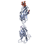
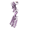
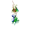
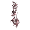
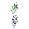
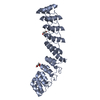
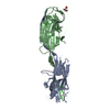
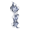
| Title | Crystal structure of C-terminal fragment of pilus adhesin SpaC from Lactobacillus rhamnosus GG |
|---|
 Components Components | Pilus assembly protein |
|---|
 Keywords Keywords | CELL ADHESION / Pilus adhesin / Tip pilin / SpaCBA pilus / sortase / Lactobacillus rhamnosus GG / pili / fimbria / Probiotics / SpaC / surface protein |
|---|
| Function / homology |  Function and homology information Function and homology information
Prealbumin-like fold domain / Prealbumin-like fold domain / von Willebrand factor type A domain / VWFA domain profile. / von Willebrand factor (vWF) type A domain / von Willebrand factor, type A / von Willebrand factor A-like domain superfamily / Immunoglobulin-like fold / Immunoglobulins / Immunoglobulin-like ...Prealbumin-like fold domain / Prealbumin-like fold domain / von Willebrand factor type A domain / VWFA domain profile. / von Willebrand factor (vWF) type A domain / von Willebrand factor, type A / von Willebrand factor A-like domain superfamily / Immunoglobulin-like fold / Immunoglobulins / Immunoglobulin-like / Sandwich / Mainly BetaSimilarity search - Domain/homology |
|---|
| Biological species |  Lactobacillus rhamnosus (bacteria) Lactobacillus rhamnosus (bacteria) |
|---|
| Method |  X-RAY DIFFRACTION / SYNCHROTRON / MOLECULAR REPLACEMENT / Resolution: 3.18 Å X-RAY DIFFRACTION / SYNCHROTRON / MOLECULAR REPLACEMENT / Resolution: 3.18 Å |
|---|
 Authors Authors | Kant, A. / Palva, A. / von Ossowski, I. / Krishnan, V. |
|---|
| Funding support |  India, 1items India, 1items | Organization | Grant number | Country |
|---|
| Department of Biotechnology (DBT, India) | BT/PR5891/BRB/10/1098/2012 | India |
|
|---|
 Citation Citation | Journal: J.Struct.Biol. / Year: 2020
Title: Crystal structure of lactobacillar SpaC reveals an atypical five-domain pilus tip adhesin: Exposing its substrate-binding and assembly in SpaCBA pili.
Authors: Kant, A. / Palva, A. / von Ossowski, I. / Krishnan, V. |
|---|
| History | | Deposition | Mar 18, 2020 | Deposition site: PDBJ / Processing site: PDBJ |
|---|
| Revision 1.0 | Jul 29, 2020 | Provider: repository / Type: Initial release |
|---|
| Revision 1.1 | Aug 19, 2020 | Group: Database references / Category: citation / Item: _citation.journal_volume |
|---|
| Revision 1.2 | Aug 26, 2020 | Group: Structure summary / Category: audit_author / Item: _audit_author.name |
|---|
| Revision 1.3 | Sep 9, 2020 | Group: Structure summary / Category: audit_author / Item: _audit_author.name |
|---|
| Revision 1.4 | Sep 15, 2021 | Group: Advisory / Database references ...Advisory / Database references / Derived calculations / Refinement description
Category: database_2 / pdbx_unobs_or_zero_occ_atoms ...database_2 / pdbx_unobs_or_zero_occ_atoms / pdbx_validate_close_contact / refine_hist / struct_conn
Item: _database_2.pdbx_DOI / _database_2.pdbx_database_accession / _refine_hist.d_res_low |
|---|
| Revision 1.5 | Nov 29, 2023 | Group: Data collection / Refinement description
Category: chem_comp_atom / chem_comp_bond / pdbx_initial_refinement_model |
|---|
| Revision 1.6 | Nov 6, 2024 | Group: Structure summary / Category: pdbx_entry_details / pdbx_modification_feature |
|---|
|
|---|
 Movie
Movie Controller

 Yorodumi
 Open data
 Basic information
 Structure visualization
 Downloads & links
 Other downloads


 PDBj
PDBj


 Assembly
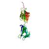
 Sample preparation
 / Beamline: ID23-1 / Wavelength: 0.87782 Å
 Processing