- PDB-6m7a: Structure of REV7-R124A complexed with SHLD3(28-73) -
+
データを開く
IDまたはキーワード:
読み込み中...
-
基本情報
登録情報
データベース: PDB / ID: 6m7a
タイトル
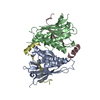
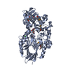
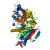
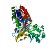
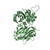
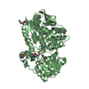
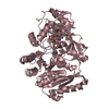
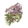
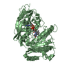
Structure of REV7-R124A complexed with SHLD3(28-73)
要素
Mitotic spindle assembly checkpoint protein MAD2B
Shieldin complex subunit 3
キーワード
REPLICATION / REV7 / shieldin / SHLD3 / DNA damage
機能・相同性
機能・相同性情報
somatic diversification of immunoglobulins involved in immune response / DNA damage response, signal transduction resulting in transcription / zeta DNA polymerase complex / positive regulation of isotype switching / negative regulation of transcription by competitive promoter binding / positive regulation of double-strand break repair via nonhomologous end joining / negative regulation of cell-cell adhesion mediated by cadherin / JUN kinase binding / negative regulation of epithelial to mesenchymal transition / negative regulation of ubiquitin protein ligase activity ...somatic diversification of immunoglobulins involved in immune response / DNA damage response, signal transduction resulting in transcription / zeta DNA polymerase complex / positive regulation of isotype switching / negative regulation of transcription by competitive promoter binding / positive regulation of double-strand break repair via nonhomologous end joining / negative regulation of cell-cell adhesion mediated by cadherin / JUN kinase binding / negative regulation of epithelial to mesenchymal transition / negative regulation of ubiquitin protein ligase activity / mitotic spindle assembly checkpoint signaling / telomere maintenance in response to DNA damage / positive regulation of peptidyl-serine phosphorylation / error-prone translesion synthesis / negative regulation of double-strand break repair via homologous recombination / Translesion synthesis by REV1 / Translesion synthesis by POLK / Translesion synthesis by POLI / actin filament organization / regulation of cell growth / negative regulation of canonical Wnt signaling pathway / negative regulation of protein catabolic process / spindle / transcription corepressor activity / site of double-strand break / double-strand break repair / chromosome / RNA polymerase II-specific DNA-binding transcription factor binding / cell division / DNA repair / chromatin / positive regulation of DNA-templated transcription / nucleolus / negative regulation of transcription by RNA polymerase II / nucleoplasm / nucleus / cytoplasm 類似検索 - 分子機能
Shieldin complex subunit 3 / Cell Cycle, Spindle Assembly Checkpoint Protein; Chain A / HORMA domain / Mad2-like / HORMA domain / HORMA domain / HORMA domain profile. / HORMA domain superfamily / 2-Layer Sandwich / Alpha Beta 類似検索 - ドメイン・相同性
 ムービー
ムービー コントローラー


 データを開く
 基本情報
基本情報 要素
要素 キーワード
キーワード 機能・相同性情報
 Homo sapiens (ヒト)
 X線回折 /
  データ登録者
 中国, 2件
  引用
引用 構造の表示
構造の表示 ダウンロードとリンク
 その他のダウンロード
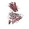

 PDBj
PDBj


 集合体
集合体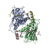

 分子量: 18.015 Da / 分子数: 381 / 由来タイプ: 天然 / 式: H2O
 試料調製
試料調製 解析
解析